
Aortic Collection
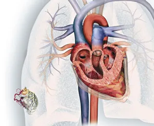
The aortic valve is a crucial component of the human heart, responsible for regulating blood flow between the left ventricle and the aorta


All Professionally Made to Order for Quick Shipping
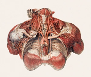
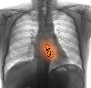
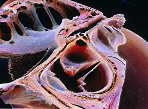
The aortic valve is a crucial component of the human heart, responsible for regulating blood flow between the left ventricle and the aorta. In this captivating cross-section image of the heart, we can see the intricate network of blood vessels that make up the coronary system. Located in the bottom left corner, it showcases not only the aorta but also coronary arteries, veins, and other blood vessels. Moving on to Picture No. 11675562, we are presented with an astonishing view of blood vessels in both chest and neck regions. The complexity and interconnectedness of these pathways highlight how vital they are for delivering oxygenated blood throughout our bodies. As we delve deeper into this series of images (Picture No. 11675561 to Picture No. 11675554), each one offers us a unique perspective on different aspects related to "aortic. " Whether it's capturing specific angles or focusing on prosthetic heart valves through X-ray technology (as seen in Prosthetic Heart Valves - X-ray F008 / 3450), these visuals provide invaluable insights into cardiac health. These pictures serve as powerful reminders of how remarkable our cardiovascular system truly is – constantly working behind-the-scenes to keep us alive and thriving. Understanding its intricacies helps medical professionals diagnose conditions such as aortic stenosis or regurgitation more accurately while guiding them towards effective treatment options. These captivating images shed light on various elements associated with "aortic, " from its role within the human heart to its connection with other crucial components like coronary arteries and veins. They remind us just how extraordinary our bodies are and emphasize why ongoing research into cardiovascular health remains essential for enhancing our understanding and improving patient care worldwide.