Fraud Blocker Skip to main content

Anatomy Collection (page 20)

"Unveiling the Intricacies of Anatomy: From Sensory Homunculus to Motor Homunculus" Step into the fascinating world of anatomy, where every detail tells a story

Background imageAnatomy Collection: Nerves of the skull and eye. Lithograph by Davesne, based on a drawing by Leveille

Nerves of the skull and eye. Lithograph by Davesne, based on a drawing by Leveille
FLO4672497 Nerves of the skull and eye. Lithograph by Davesne, based on a drawing by Leveille, in Petity Atlas complet d'Anatomie descriptive du Corps Humain, by Dr

Background imageAnatomy Collection: Ophthalmic and maxillary nerves of the fifth cranial nerve. Lithograph by Davesne

Ophthalmic and maxillary nerves of the fifth cranial nerve. Lithograph by Davesne, based on a drawing by Leveille
FLO4672496 Ophthalmic and maxillary nerves of the fifth cranial nerve. Lithograph by Davesne, based on a drawing by Leveille, in Petity Atlas complet d'Anatomie descriptive du Corps Humain

Background imageAnatomy Collection: Lumbar plexus and crural nerve. Lithograph of Corbie, based on a drawing by Leveille

Lumbar plexus and crural nerve. Lithograph of Corbie, based on a drawing by Leveille
FLO4672455 Lumbar plexus and crural nerve. Lithograph of Corbie, based on a drawing by Leveille, in Petity Atlas complet d'Anatomie descriptive du Corps Humain, by Dr

Background imageAnatomy Collection: Shoulder and arm bone: scapula, humerus, cubitus and radius. Lithograph of Corbie

Shoulder and arm bone: scapula, humerus, cubitus and radius. Lithograph of Corbie
FLO4672433 Shoulder and arm bone: scapula, humerus, cubitus and radius. Lithograph of Corbie, in Petity Atlas complet d'Anatomie descriptive du Corps Humain, by Dr

Background imageAnatomy Collection: Side view of human skeleton and horse skeleton. Copperplate engraving by Edward Mitchell after

Side view of human skeleton and horse skeleton. Copperplate engraving by Edward Mitchell after anatomical illustrations
FLO4565764 Side view of human skeleton and horse skeleton. Copperplate engraving by Edward Mitchell after anatomical illustrations by Bernhard Siegfried Albinus

Background imageAnatomy Collection: Fetal skeleton at six months with scythe 1, at three months 2 and at ten weeks 3

Fetal skeleton at six months with scythe 1, at three months 2 and at ten weeks 3
FLO4565763 Fetal skeleton at six months with scythe 1, at three months 2 and at ten weeks 3. Copperplate engraving by Edward Mitchell after an anatomical illustration by Jean-Joseph Sue from John

Background imageAnatomy Collection: Fetal skeleton at the time of birth. Copperplate engraving by Edward Mitchell after an anatomical

Fetal skeleton at the time of birth. Copperplate engraving by Edward Mitchell after an anatomical illustration by
FLO4565750 Fetal skeleton at the time of birth. Copperplate engraving by Edward Mitchell after an anatomical illustration by Jean-Joseph Sue from John Barclay's A Series of Engravings of

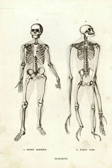
Background imageAnatomy Collection: Skeletons of a man, Homo sapiens, and endangered lar gibbon, Hylobates lar (Simia lar)

Skeletons of a man, Homo sapiens, and endangered lar gibbon, Hylobates lar (Simia lar)
FLO4720006 Skeletons of a man, Homo sapiens, and endangered lar gibbon, Hylobates lar (Simia lar). Lithograph from Georg Friedrich Treitschke's Gallery of Natural History

Background imageAnatomy Collection: Asthma. Engraving from ' Les Grands Ales et les Grands Remedes' by Jules Rengade, 1889

Asthma. Engraving from " Les Grands Ales et les Grands Remedes" by Jules Rengade, 1889
PCT4274721 Asthma. Engraving from " Les Grands Ales et les Grands Remedes" by Jules Rengade, 1889.; (add.info.: Asthma)

Background imageAnatomy Collection: Jan Evangelista Purkyne, 19th century (engraving)

Jan Evangelista Purkyne, 19th century (engraving)
3733543 Jan Evangelista Purkyne, 19th century (engraving); (add.info.: Portrait of Jan Evangelista Purkyne (1787-1869) a Czech anatomist and physiologist

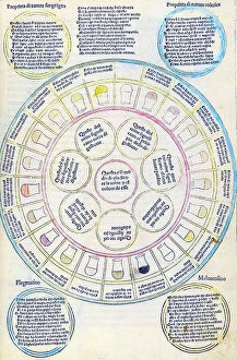
Background imageAnatomy Collection: The Canon of Medicine by Avicenna

The Canon of Medicine by Avicenna
2625213 The Canon of Medicine by Avicenna; (add.info.: The central text consists of the anatomical sections of the Canon of Medicine by Avicenna)

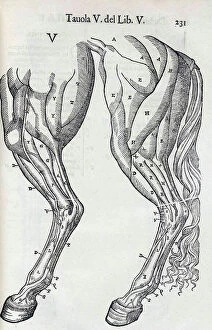
Background imageAnatomy Collection: Equine anatomical Illustration

Equine anatomical Illustration
2626318 Equine anatomical Illustration; (add.info.: Equine anatomical Illustration from 'Anatomia del cavallo, infermita, et suoi rimedii')

Background imageAnatomy Collection: Equine anatomical Illustration

Equine anatomical Illustration
2626316 Equine anatomical Illustration; (add.info.: Equine anatomical Illustration from 'Anatomia del cavallo, infermita, et suoi rimedii')

Background imageAnatomy Collection: Equine anatomical Illustration

Equine anatomical Illustration
2626314 Equine anatomical Illustration; (add.info.: Equine anatomical Illustration from 'Anatomia del cavallo, infermita, et suoi rimedii')

Background imageAnatomy Collection: Equine anatomical Illustration

Equine anatomical Illustration
2626312 Equine anatomical Illustration; (add.info.: Equine anatomical Illustration from 'Anatomia del cavallo, infermita, et suoi rimedii')

Background imageAnatomy Collection: Shi si jing fa hui

Shi si jing fa hui
2626543 Shi si jing fa hui; (add.info.: Illustration from the Japanese & Chinese edition (1716) of 'Shi si jing fa hui'

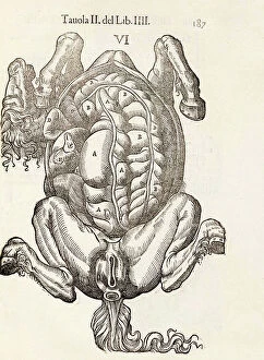
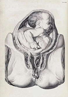
Background imageAnatomy Collection: A set of anatomical tables, with explanations, and an abridgment, of the practice of midwifery

A set of anatomical tables, with explanations, and an abridgment, of the practice of midwifery, 18th (engraving)
2626224 A set of anatomical tables, with explanations, and an abridgment, of the practice of midwifery, 18th (engraving); (add.info)

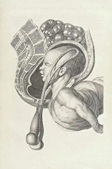
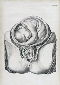
Background imageAnatomy Collection: A set of anatomical tables, with explanations, and an abridgment, of the practice of midwifery

A set of anatomical tables, with explanations, and an abridgment, of the practice of midwifery, 18th (engraving)
2626223 A set of anatomical tables, with explanations, and an abridgment, of the practice of midwifery, 18th (engraving); (add.info)

Background imageAnatomy Collection: A set of anatomical tables, with explanations, and an abridgment, of the practice of midwifery

A set of anatomical tables, with explanations, and an abridgment, of the practice of midwifery, 18th (engraving)
2626220 A set of anatomical tables, with explanations, and an abridgment, of the practice of midwifery, 18th (engraving); (add.info)

Background imageAnatomy Collection: Fasciculus medicinae

Fasciculus medicinae
2626474 Fasciculus medicinae; (add.info.: Illustration from 'Fasciculus medicinae' Venice; 1494 the first printed book to contain anatomical illustrations)

Background imageAnatomy Collection: Les oeuures d'Ambroise Pare?

Les oeuures d'Ambroise Pare?
2626406 Les oeuures d'Ambroise Pare?; (add.info.: Illustration from 'Les oeuures d'Ambroise Pare?' 1585 by Ambroise Pare)

Background imageAnatomy Collection: Shi si jing fa hui

Shi si jing fa hui
2626558 Shi si jing fa hui; (add.info.: Illustration from the Japanese & Chinese edition (1716) of 'Shi si jing fa hui'

Background imageAnatomy Collection: Equine anatomical Illustration

Equine anatomical Illustration
2626311 Equine anatomical Illustration; (add.info.: Equine anatomical Illustration from 'Anatomia del cavallo, infermita, et suoi rimedii')

Background imageAnatomy Collection: A set of anatomical tables, with explanations, and an abridgment, of the practice of midwifery

A set of anatomical tables, with explanations, and an abridgment, of the practice of midwifery, 18th (engraving)
2626256 A set of anatomical tables, with explanations, and an abridgment, of the practice of midwifery, 18th (engraving); (add.info)

Background imageAnatomy Collection: Shi si jing fa hui

Shi si jing fa hui
2626540 Shi si jing fa hui; (add.info.: Illustration from the Japanese & Chinese edition (1716) of 'Shi si jing fa hui'

Background imageAnatomy Collection: A set of anatomical tables, with explanations, and an abridgment, of the practice of midwifery

A set of anatomical tables, with explanations, and an abridgment, of the practice of midwifery, 18th (engraving)
2626219 A set of anatomical tables, with explanations, and an abridgment, of the practice of midwifery, 18th (engraving); (add.info)

Background imageAnatomy Collection: The anatomy textbook 'Ontleding des Menschelyken Lichaams', 1690 (engraving)

The anatomy textbook 'Ontleding des Menschelyken Lichaams', 1690 (engraving)
2625813 The anatomy textbook 'Ontleding des Menschelyken Lichaams', 1690 (engraving); (add.info.: Illustrations by Govard Bidloo)

Background imageAnatomy Collection: Les oeuures d'Ambroise Pare?

Les oeuures d'Ambroise Pare?
2626404 Les oeuures d'Ambroise Pare?; (add.info.: Illustration from 'Les oeuures d'Ambroise Pare?' 1585 by Ambroise Pare)

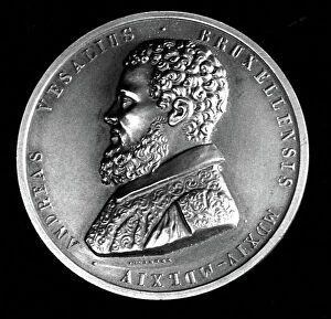
Background imageAnatomy Collection: Medal depicting Andreas Vesalius

Medal depicting Andreas Vesalius
3586027 Medal depicting Andreas Vesalius; (add.info.: Medal depicting Andreas Vesalius (1514-1564) a Flemish/Netherlandish anatomist, physician, and author of books on human anatomy

Background imageAnatomy Collection: William Henry Flower (1831-1899)

William Henry Flower (1831-1899)
865073 William Henry Flower (1831-1899); (add.info.: William Henry Flower (1831-1899) English surgeon and zoologist, born at Stratford-on-Avon, Warwickshire, England

Background imageAnatomy Collection: Internal organs of the human body. From Gregor Reisch Margarita Philosophica, Basle, 1508

Internal organs of the human body. From Gregor Reisch Margarita Philosophica, Basle, 1508
527495 Internal organs of the human body. From Gregor Reisch Margarita Philosophica, Basle, 1508. ; Universal History Archive/UIG.

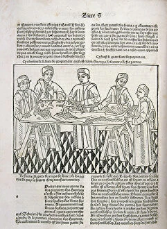
Background imageAnatomy Collection: A dissection in progress. From Le livre de la propriete des choses (On the Properites of Things)

A dissection in progress. From Le livre de la propriete des choses (On the Properites of Things)
527490 A dissection in progress. From Le livre de la propriete des choses (On the Properites of Things) written by the English Franciscan monk Bartholomew Glanville, called Bartolomaeus Anglicus

Background imageAnatomy Collection: Nerves involved in the sense of taste (coloured engraving)

Nerves involved in the sense of taste (coloured engraving)
8655717 Nerves involved in the sense of taste (coloured engraving) by French School, (19th century) (after); Private Collection; (add.info.: Nerves involved in the sense of taste)

Background imageAnatomy Collection: Cross-section of the skin (coloured engraving)

Cross-section of the skin (coloured engraving)
8655716 Cross-section of the skin (coloured engraving) by French School, (19th century) (after); Private Collection; (add.info.: Cross-section of the skin)

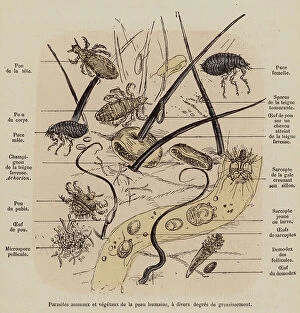
Background imageAnatomy Collection: Parasites of the human skin (coloured engraving)

Parasites of the human skin (coloured engraving)
8655715 Parasites of the human skin (coloured engraving) by French School, (19th century) (after); Private Collection; (add.info.: Parasites of the human skin)

Background imageAnatomy Collection: Muscular system of the human body (coloured engraving)

Muscular system of the human body (coloured engraving)
8655713 Muscular system of the human body (coloured engraving) by French School, (19th century) (after); Private Collection; (add.info.: Muscular system of the human body (front view)

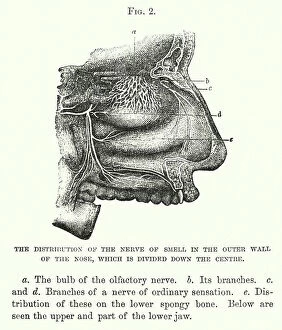
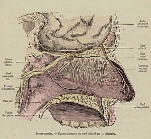
Background imageAnatomy Collection: The distribution of the nerve of smell in the outer wall of the nose

The distribution of the nerve of smell in the outer wall of the nose, which is divided down the centre (engraving)
8646012 The distribution of the nerve of smell in the outer wall of the nose, which is divided down the centre (engraving) by English School

Background imageAnatomy Collection: Glottis and vocal cords (engraving)

Glottis and vocal cords (engraving)
8655777 Glottis and vocal cords (engraving) by French School, (19th century) (after); Private Collection; (add.info.: Glottis and vocal cords)

Background imageAnatomy Collection: Oxygenation of blood in the lungs (coloured engraving)

Oxygenation of blood in the lungs (coloured engraving)
8655756 Oxygenation of blood in the lungs (coloured engraving) by French School, (19th century) (after); Private Collection; (add.info.: Oxygenation of blood in the lungs)

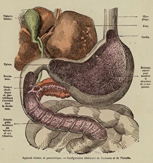
Background imageAnatomy Collection: Organs of the digestive system (coloured engraving)

Organs of the digestive system (coloured engraving)
8655752 Organs of the digestive system (coloured engraving) by French School, (19th century) (after); Private Collection; (add.info.: Organs of the digestive system)

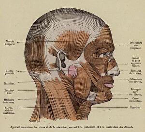
Background imageAnatomy Collection: Muscles of the jaw and lips and their role in eating food (coloured engraving)

Muscles of the jaw and lips and their role in eating food (coloured engraving)
8655748 Muscles of the jaw and lips and their role in eating food (coloured engraving) by French School, (19th century) (after); Private Collection; (add.info)

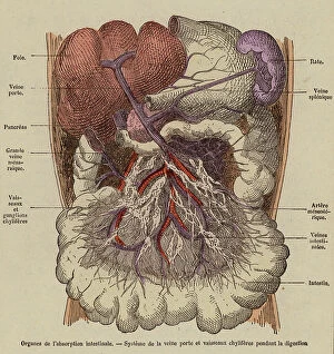
Background imageAnatomy Collection: Organs of absorption in the digestive system (coloured engraving)

Organs of absorption in the digestive system (coloured engraving)
8655745 Organs of absorption in the digestive system (coloured engraving) by French School, (19th century) (after); Private Collection; (add.info.: Organs of absorption in the digestive system)

Background imageAnatomy Collection: Intestinal villi (coloured engraving)

Intestinal villi (coloured engraving)
8655743 Intestinal villi (coloured engraving) by French School, (19th century) (after); Private Collection; (add.info.: Intestinal villi)

Background imageAnatomy Collection: Cross-section of the neck at the leval of the sixth cervical vertebra (coloured engraving)

Cross-section of the neck at the leval of the sixth cervical vertebra (coloured engraving)
8655734 Cross-section of the neck at the leval of the sixth cervical vertebra (coloured engraving) by French School, (19th century) (after); Private Collection; (add.info)

Background imageAnatomy Collection: Brain seen from below (coloured engraving)

Brain seen from below (coloured engraving)
8655729 Brain seen from below (coloured engraving) by French School, (19th century) (after); Private Collection; (add.info.: Brain seen from below)

Background imageAnatomy Collection: Section of the spinal cord showing the roots of the spinal nerves (coloured engraving)

Section of the spinal cord showing the roots of the spinal nerves (coloured engraving)
8655727 Section of the spinal cord showing the roots of the spinal nerves (coloured engraving) by French School, (19th century) (after); Private Collection; (add.info)

Background imageAnatomy Collection: Nerves of the arm and leg (coloured engraving)

Nerves of the arm and leg (coloured engraving)
8655725 Nerves of the arm and leg (coloured engraving) by French School, (19th century) (after); Private Collection; (add.info.: Nerves of the arm and leg)

Background imageAnatomy Collection: Nasal cavity and olfactory tract (coloured engraving)

Nasal cavity and olfactory tract (coloured engraving)
8655719 Nasal cavity and olfactory tract (coloured engraving) by French School, (19th century) (after); Private Collection; (add.info.: Nasal cavity and olfactory tract)


All Professionally Made to Order for Quick Shipping

"Unveiling the Intricacies of Anatomy: From Sensory Homunculus to Motor Homunculus" Step into the fascinating world of anatomy, where every detail tells a story. Explore the depths of human and animal structures as we delve into this captivating subject. Let's begin with Leonardo da Vinci's masterpiece, "Head of a Bear. " Created in 1480 but discovered in 1945, this artwork showcases da Vinci's unparalleled ability to capture anatomical accuracy. The intricate details reveal his keen observation skills and deep understanding of form. Moving on to our furry friends, the greyhound takes center stage. Discovering the anatomy of these majestic creatures unveils their remarkable speed and agility. From their sleek skeleton to their muscular build, it is evident why they are renowned for their racing abilities. But what about us humans? Enter the sensory homunculus – a visual representation that depicts how our brain perceives different body parts based on sensitivity levels. This map reveals intriguing insights into how our senses are distributed throughout our bodies. X-ray images provide another dimension to understanding anatomy. Take a glimpse at normal knees through an X-ray lens – marvel at the complexity hidden beneath our skin while appreciating its delicate balance between strength and flexibility. The backbone is often considered one of nature's most ingenious designs. Our human backbone includes ribs and pelvis, providing stability while allowing movement – truly an architectural marvel worth exploring further. Venturing deeper within ourselves, let us explore the intricacies of the human brain from an inferior view. Witness its complex network of connections responsible for controlling various bodily functions - truly awe-inspiring. As we journey back in time, we encounter hominid crania – remnants that shed light on our evolutionary history. These ancient skulls offer glimpses into early forms such as Australopithecus afarensis (AL 288-1), famously known as Lucy - bridging gaps between past and present.