Fraud Blocker Skip to main content

Algal Collection (page 6)

"Exploring the Intricate World Beauty" Immerse yourself in the captivating artistry of diatom algae, as depicted by Ernst Haeckel

Background imageAlgal Collection: Chordaria flagellifornis, seaweed

Chordaria flagellifornis, seaweed
Plate 57 from Algae Danmonienses : or dried specimens of Marine Plants, principally collected in Devonshire by Mary Wyatt. 1834-1840

Background imageAlgal Collection: Asperoccus turneri, seaweed

Asperoccus turneri, seaweed
Plate 59 from Algae Danmonienses : or dried specimens of Marine Plants, principally collected in Devonshire by Mary Wyatt. 1834-1840

Background imageAlgal Collection: Fucus agarum, kelp

Fucus agarum, kelp
Plate 75 from Fuci, or coloured figures and descriptions of the Plants referred by botanists to the genus Fucus (1808-1819), Volume II, by Mary Dawson Turner

Background imageAlgal Collection: Chondus cripus, carrageen moss

Chondus cripus, carrageen moss
Illustration from Algae Danmonienses: or dried specimens of Marine Plants, principally collected in Devonshire by Mary Wyatt; carefully named according to Dr. Hookers British Flora

Background imageAlgal Collection: Rhodophyta, Coralline algae

Rhodophyta, Coralline algae
This specimen was collected by Charles Darwin in 1836 from Keeling Atoll, Indian Ocean

Background imageAlgal Collection: Raphiidonema faringdone, calcareous sponge

Raphiidonema faringdone, calcareous sponge
A vase-shaped calcareous sponge with numerous small canals from the Cretaceous of Berkshire, England

Background imageAlgal Collection: Chleophaga hybrida, kelp goose

Chleophaga hybrida, kelp goose
Ff. 66. Watercolour painting by George Forster annotated Anas ganta Anas antarctica and made during Captain James Cooks second voyage to explore the southern continent (1772-75)

Background imageAlgal Collection: Coccoliths magnified a thousand times

Coccoliths magnified a thousand times
An illustration of Coccoliths magnified a thousand times. Coccoliths are micro-fossils and feature heavily in the composition of chalk

Background imageAlgal Collection: Lichens

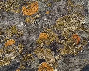
Lichens result from an intimate relationship between a fungus and an alga; there are about 18, 000 species

Background imageAlgal Collection: Calcidiscus leptoporus and Syracolithus quadriperforatus, co

Calcidiscus leptoporus and Syracolithus quadriperforatus, co
In this scanning electron micrograph, the transition of a life-cycle stage in Calcidiscus is shown from the outer cover to the inner layer. Specimen taken from W. Mediterranean

Background imageAlgal Collection: Actinoptychus, diatom

Actinoptychus, diatom
Scanning electron microscope image of the exterior valve of the diatom Actinoptychus (x 500 on a standard 9 cm wide print)

Background imageAlgal Collection: Chalk

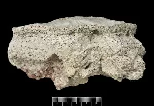
Chalk
A piece of flintless white chalk from the Upper Chalk, Flamborough, Yorks. Chalk is a sedimentary rock formed in deep seas

Background imageAlgal Collection: Marpolia spissa, fossilised algae

Marpolia spissa, fossilised algae
A fossilised type of green algae, possible filamentous cyanobacteria (Marpolia spissa), approximately 12 mm in height. This specimen was discovered in the Middle Cambrian, Burgess Shale, Canada

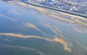
Background imageAlgal Collection: Orange Algae, aerial view of bloom floating on surface of sea, Morecambe Bay, Blackpool

Orange Algae, aerial view of bloom floating on surface of sea, Morecambe Bay, Blackpool, Lancashire, England, August

Background imageAlgal Collection: Blue-green Algae (Rivularia bullata) Growing amid and on Acorn Barnacles / Padstow, Cornwall

Blue-green Algae (Rivularia bullata) Growing amid and on Acorn Barnacles / Padstow, Cornwall

Background imageAlgal Collection: Green Alga (Trentepohlia aurea) orange filamentous alga growing on old gravestone in churchyard

Green Alga (Trentepohlia aurea) orange filamentous alga growing on old gravestone in churchyard, Culbone, Exmoor, Somerset, England, march

Background imageAlgal Collection: RedAlgae (Porphyridium cruentum)

RedAlgae (Porphyridium cruentum)

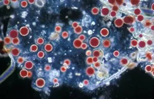
Background imageAlgal Collection: Pl ton -(Noctiluca scintillans) group / one with ingested copepod / x12

Pl ton -(Noctiluca scintillans) group / one with ingested copepod / x12

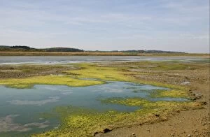
Background imageAlgal Collection: View of algae on lagoon in coastal marshland habitat, Cley Marshes Reserve, Cley-next-the-sea

View of algae on lagoon in coastal marshland habitat, Cley Marshes Reserve, Cley-next-the-sea, Norfolk, England, april

Background imageAlgal Collection: Green algae on pond surface, Stowmarket, Suffolk, England, april

Green algae on pond surface, Stowmarket, Suffolk, England, april

Background imageAlgal Collection: Red Cyanobacteria (Cyanobacteria sp. ) forming scum on pool in freshwater marsh, Ceredigion

Red Cyanobacteria (Cyanobacteria sp. ) forming scum on pool in freshwater marsh, Ceredigion, Wales, june
Red Cyanobacteria (Cyanobacteria sp.) forming scum on pool in freshwater marsh, Ceredigion, Wales, june

Background imageAlgal Collection: Common Kestrel (Falco tinnunculus) immature male, first summer plumage

Common Kestrel (Falco tinnunculus) immature male, first summer plumage, landing on algal mat in pond, Norfolk, England, april

Background imageAlgal Collection: Calcareous phytoplankton fossil, artwork

Calcareous phytoplankton fossil, artwork
Calcareous phytoplankton fossil. Artwork of a fossil of the skeleton (coccosphere) of a coccolithotrope, a small marine algal organism

Background imageAlgal Collection: Plankton cell wall, SEM

Plankton cell wall, SEM
Plankton cell wall. Coloured scanning electron micrograph (SEM) of the geometrical plates making up the mineralised cell wall of a planktonic alga (Coronosphaera mediterranea)

Background imageAlgal Collection: Calcareous phytoplankton plates, SEM

Calcareous phytoplankton plates, SEM
Calcareous phytoplankton plates. Coloured scanning electron micrograph (SEM) of parts of the skeleton (coccosphere) of the coccolithophore Emiliania huxleyi, a small marine algal organism

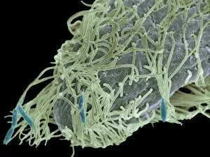
Background imageAlgal Collection: Alga-covered protozoan, SEM

Alga-covered protozoan, SEM
Alga-covered protozoan, coloured scanning electron micrograph (SEM). Algae (blue) are seen on the body of the protozoan, and cilia (green) are used to waft the algae to the protozoans feeding area

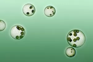
Background imageAlgal Collection: Green algae colonies

Green algae colonies. Light micrograph of green algae colonies (Volvox sp.). These colonies are made up of approximately 500 individual flagellate cells, arranged in a glycoprotein filled sphere

Background imageAlgal Collection: Rhabdonema marine diatom, SEM

Rhabdonema marine diatom, SEM
Rhabdonema. Coloured scanning electron micrograph (SEM) of a colony of the marine diatom Rhabdonema sp. Magnification: x500 when printed at 10 centimetres wide

Background imageAlgal Collection: Licmorpha freshwater diatom, SEM

Licmorpha freshwater diatom, SEM
Licmorpha. Coloured scanning electron micrograph (SEM) of colonies of the freshwater diatom Licmorpha sp. Magnification: x500 when printed at 10 centimetres wide

Background imageAlgal Collection: Algal bloom

Algal bloom, aerial photograph. The red colour of the water is caused by the proliferation of microscopic photosynthetic organisms in the seawater

Background imageAlgal Collection: Worm feeding on algae, light micrograph

Worm feeding on algae, light micrograph
Worm feeding on algae. Light micrograph of an microscopic annelid worm (subclass Oligochaeta, upper left) feeding on green algae

Background imageAlgal Collection: Microsterias green alga, light micrograph

Microsterias green alga, light micrograph
Microsterias green alga. Light micrograph of a Microsterias sp. desmid green algae. Desmids are a common group of unicellular freshwater algae that have intricate cell walls

Background imageAlgal Collection: Algae

Algae. Photographed in the Red Sea

Background imageAlgal Collection: Pediastrum green algae, light micrograph

Pediastrum green algae, light micrograph
Pediastrum green algae. Light micrograph of a Pediastrum sp. green alga. Members of this genus are non-motile algae that live in freshwater environments

Background imageAlgal Collection: Morning Glory Pool, Yellowstone

Morning Glory Pool, Yellowstone National Park, Wyoming, USA. This geothermal pool is warmed by the volcanic activity beneath it

Background imageAlgal Collection: Epistylis protozoan colony on algae

Epistylis protozoan colony on algae. Light micrograph of a colony of Epistylis sp. protozoa (centre right) attached to a green algae filament (lower left)

Background imageAlgal Collection: Diatoms, light micrograph

Diatoms, light micrograph. Diatoms are a group of photosynthetic, single-celled algae containing about 10, 000 species. They form an important part of the plankton at the base of the marine

Background imageAlgal Collection: Melosira sp diatoms, light micrograph

Melosira sp diatoms, light micrograph
Diatoms. Light micrograph of Melosira monoliformis freshwater diatoms. Diatoms are a group of photosynthetic, single-celled algae containing about 10, 000 species

Background imageAlgal Collection: Pleurosigma sp diatoms, light micrograph

Pleurosigma sp diatoms, light micrograph
Diatoms. Differential interference contrast micrograph of Pleurosigma angulatum marine diatoms. Diatoms are a group of photosynthetic, single-celled algae containing about 10, 000 species

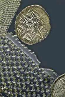
Background imageAlgal Collection: Fossil diatoms, light micrograph

Fossil diatoms, light micrograph. Diatoms are a group of photosynthetic, single-celled algae containing about 10, 000 species


All Professionally Made to Order for Quick Shipping

"Exploring the Intricate World Beauty" Immerse yourself in the captivating artistry of diatom algae, as depicted by Ernst Haeckel. These microscopic organisms showcase their intricate structures and mesmerizing patterns, reminding us of the boundless creativity found in nature. Pressed seaweed specimens C016 / 6127 offer a glimpse into the diverse forms and textures that algal species can take. From delicate filaments to robust fronds, each specimen tells a unique story of adaptation and survival in marine ecosystems. Behold the beauty of calcareous phytoplankton through scanning electron microscopy (SEM). The exquisite details captured reveal their ornate shells and elaborate architecture, serving as a testament to their vital role in oceanic food webs. Discosphaera tubifera, a coccolithophore, enchants with its spherical shape adorned by intricate calcium carbonate plates. This tiny organism plays an essential part in carbon cycling and contributes to the stunning white cliffs seen along some coastlines. Dictyota dichotoma showcases its elegant branching structure, exemplifying how algae can create complex habitats for other marine organisms. Its presence enriches coastal ecosystems while providing shelter for countless creatures. Diatoms take center stage once again under SEM's magnifying lens. Their symmetrical siliceous skeletons exhibit remarkable diversity – from star-shaped patterns to delicate lace-like designs – showcasing nature's endless ingenuity. Marvel at Fucus bulbosus or kelp's majestic presence as it sways gracefully beneath ocean currents. These large brown algae provide refuge for numerous marine species while contributing to nutrient cycling within coastal environments. Microcystis blue-green alga captivates with its vibrant hue amidst freshwater bodies. Though often associated with harmful algal blooms, this organism also serves ecological functions such as nitrogen fixation and oxygen production. Witness yet another diatom masterpiece through SEM imagery – their intricately sculpted shapes and delicate ornamentation never cease to amaze.