
3 D Visualization Collection
"Unlocking the Invisible: Exploring the World of 3D Visualization in Virology" Delve into a realm where viruses come to life through stunning computer models


All Professionally Made to Order for Quick Shipping
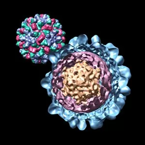
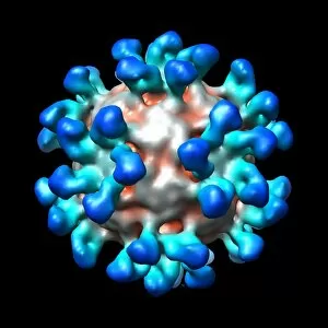
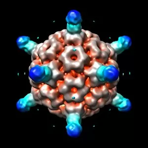
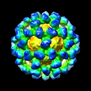
"Unlocking the Invisible: Exploring the World of 3D Visualization in Virology" Delve into a realm where viruses come to life through stunning computer models. Witness the intricate beauty and complexity of Bacteriophage phi29, as it reveals its structural secrets in an awe-inspiring visualization. Step further into this virtual world, where Simian immunodeficiency virus (SIV) takes center stage. Unravel its hidden mysteries and gain a deeper understanding of its structure and behavior. Marvel at the captivating dance between Murine norovirus and antibody fragments, as they intertwine in a mesmerizing display of molecular interactions. Embark on a journey with Bacteriophage phi29 once again, but this time through an intricately detailed computer model that showcases every minute detail with astonishing precision. Witness Cucumber necrosis virus come alive before your eyes, as it unveils itself through an immersive digital representation that captures its essence flawlessly. Explore Human rhinovirus like never before, as a meticulously crafted computer model brings forth its complex architecture and offers insights into potential treatment strategies. Be captivated by Ribgrass mosaic virus's elegant form taking shape within a virtual landscape, allowing us to study its structure from every angle imaginable. Experience the power of antibodies against Human rhinovirus firsthand through an extraordinary visual narrative that highlights their crucial role in neutralizing viral threats. Immerse yourself once more in the world of Bacteriophage P22 through an exquisitely detailed computer model that unravels its inner workings with unparalleled clarity. Witness Cucumber necrosis virus reveal itself yet again, this time accompanied by antibodies that provide vital defense mechanisms against infection - a testament to nature's ingenuity. Finally, marvel at Cucumber mosaic virus as it materializes within our screens via cutting-edge technology. This vivid depiction allows scientists to delve deeper into understanding how it operates and explore potential avenues for intervention.