Canvas Print : Nasal cavity, SEM


![]()
Canvas Prints from Science Photo Library
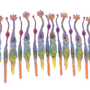
Nasal cavity, SEM
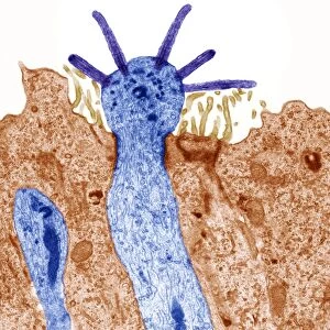
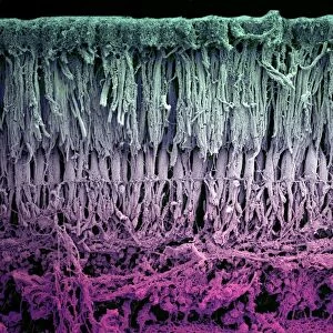
Nasal cavity. Coloured scanning electron micrograph (SEM) of the surface of the nasal cavity. It is covered in epithelial hair-like structures known as cilia (orange), which line the respiratory tract. They are covered with a wet, sticky mucus that humidifies inhaled air and traps dust particles and other air pollutants. Co-ordinated, wave-like beating of the cilia propels the mucus to the back of the nose (pharynx), where it is swallowed. Magnification: x1800 at 6x7cm size
Science Photo Library features Science and Medical images including photos and illustrations
Media ID 6422248
© SUSUMU NISHINAGA/SCIENCE PHOTO LIBRARY
Cilia Ciliated Cilium Epidermal Epidermis Hair Hairs Hairy Mucus Nasal Cavity Nose Olfaction Respiratory Tract Sensory Smell
20"x16" (50x40cm) Canvas Print
Discover the intricacies of the human body with Media Storehouse's Canvas Prints. This captivating SEM image from Science Photo Library showcases the complex structure of the nasal cavity. Coloured in high definition, the print reveals the intricate details of the epithelial hairs, or cilia (orange), that line the nasal cavity, playing a crucial role in filtering and warming the air we breathe. Bring this stunning piece of scientific art into your home or office to ignite curiosity and inspire new perspectives.
Ready to hang Premium Gloss Canvas Print. Our archival quality canvas prints are made from Polyester and Cotton mix and stretched over a 1.25" (32mm) kiln dried knot free wood stretcher bar. Packaged in a plastic bag and secured to a cardboard insert for transit.
Canvas Prints add colour, depth and texture to any space. Professionally Stretched Canvas over a hidden Wooden Box Frame and Ready to Hang
Estimated Product Size is 40.6cm x 50.8cm (16" x 20")
These are individually made so all sizes are approximate
Artwork printed orientated as per the preview above, with portrait (vertical) orientation to match the source image.
EDITORS COMMENTS
This print from Science Photo Library offers a mesmerizing glimpse into the intricate world of our nasal cavity. In this colored scanning electron micrograph (SEM), we are presented with a close-up view of the surface of the nasal cavity, revealing an astonishingly complex network of tiny structures. The focal point of this image is undoubtedly the vibrant orange cilia that densely cover the respiratory tract lining. These hair-like structures play a crucial role in maintaining our respiratory health by performing multiple functions simultaneously. Not only do they act as sensory receptors for our olfactory sense, enabling us to perceive various scents and smells, but they also serve as guardians against harmful air pollutants. Coordinated in their movements, these cilia beat rhythmically in wave-like motions to propel a wet and sticky mucus towards the back of our nose or pharynx. This process not only humidifies inhaled air but also effectively traps dust particles and other airborne contaminants before being swallowed. At a magnification level of x1800 on a 6x7cm size scale, this SEM photograph showcases the remarkable complexity hidden within such seemingly ordinary body parts. It reminds us once again how intricately designed and interconnected every aspect of our human body truly is.
MADE IN THE UK
Safe Shipping with 30 Day Money Back Guarantee
FREE PERSONALISATION*
We are proud to offer a range of customisation features including Personalised Captions, Color Filters and Picture Zoom Tools
SECURE PAYMENTS
We happily accept a wide range of payment options so you can pay for the things you need in the way that is most convenient for you
* Options may vary by product and licensing agreement. Zoomed Pictures can be adjusted in the Basket.