Antique Framed Print : 14th Century depiction of dissection


![]()
Framed Photos from Science Photo Library
14th Century depiction of dissection
Human dissection, 14th Century style. This image is the earliest known representation of a dissection taking place. The female corpse (right) has had most of her internal organs removed. At top right are her lungs and kidneys, whilst at bottom right are her heart and stomach. The physicians assistant (centre) is holding the womans liver. The physician himself is directing his assistant, whilst a monk (far left) looks on
Science Photo Library features Science and Medical images including photos and illustrations
Media ID 6432601
© SCIENCE PHOTO LIBRARY
14th Century Dissection Historical Image Imagery Physician Female Anatomy
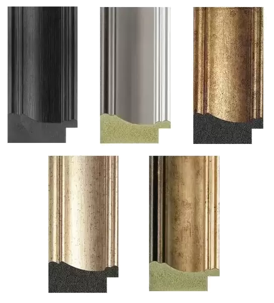
14"x12" (36x31cm) Antique Frame
Bevelled wood effect frame, card mounted, 10x8 archival quality photo print. Overall outside dimensions 14x12 inches (36x31cm). Environmentally and ozone friendly, the Polycore® moulding has the look of real wood, is durable and light and easy to hang. Biodegradable and made with non-chlorinated gases (no toxic fumes) it is efficient; producing 100 tons of polystyrene can save 300 tons of trees! Prints are glazed with lightweight, shatterproof, optical clarity acrylic (providing the same general protection from the environment as glass). The back is stapled hardboard with a sawtooth hanger attached. Note: To minimise original artwork cropping, for optimum layout, and to ensure print is secure, the visible print may be marginally smaller
Bevelled Wood Effect Framed and Mounted Prints - Professionally Made and Ready to Hang
Estimated Image Size (if not cropped) is 24.4cm x 18.1cm (9.6" x 7.1")
Estimated Product Size is 36.3cm x 31.2cm (14.3" x 12.3")
These are individually made so all sizes are approximate
Artwork printed orientated as per the preview above, with landscape (horizontal) orientation to match the source image.
EDITORS COMMENTS
This intriguing print from Science Photo Library showcases a remarkable 14th-century depiction of dissection, offering us a glimpse into the early practices of anatomy and medicine. The image captures a pivotal moment in history, as it is the earliest known representation of an actual dissection taking place. In this scene, we see a female corpse lying on a table, her body opened up for examination. Most of her internal organs have been carefully removed, revealing an astonishing level of anatomical knowledge for its time. At the top right corner, we can observe her lungs and kidneys displayed with precision, while at the bottom right lie her heart and stomach. The focus shifts to the central figures in this composition—the physician himself and his diligent assistant holding the woman's liver. The physician directs his assistant with expertise and authority as they navigate through this groundbreaking exploration of human anatomy. Adding another layer to this historical tableau is a monk positioned on the far left side. His presence suggests that religion played a significant role in shaping attitudes towards dissection during that era. As we delve into this mesmerizing snapshot from medical history, we are reminded of how far our understanding has progressed since then. This image serves as both an invaluable record of our past and an inspiration for future advancements in science and medicine.
MADE IN THE UK
Safe Shipping with 30 Day Money Back Guarantee
FREE PERSONALISATION*
We are proud to offer a range of customisation features including Personalised Captions, Color Filters and Picture Zoom Tools
SECURE PAYMENTS
We happily accept a wide range of payment options so you can pay for the things you need in the way that is most convenient for you
* Options may vary by product and licensing agreement. Zoomed Pictures can be adjusted in the Basket.