Fraud Blocker Skip to main content

Home > All Images > 2013 > December > 18 Dec 2013

Images Dated 18th December 2013 (page 2)

102 items

Background imageImages Dated 18th December 2013: Marina Bay Arts Science Museum, Singapore, Southeast Asia, Asia

Marina Bay Arts Science Museum, Singapore, Southeast Asia, Asia

Background imageImages Dated 18th December 2013: Didinium ciliate protozoan, SEM C019 / 0235

Didinium ciliate protozoan, SEM C019 / 0235
Didinium sp. ciliate protozoan, coloured scanning electron micrograph (SEM). This tiny single-celled organism is found in freshwater and marine habitats

Background imageImages Dated 18th December 2013: Genetic karyotype analysis C019 / 0270

Genetic karyotype analysis C019 / 0270
MODEL RELEASED. Genetic karyotype analysis. Analyst in a genetics laboratory studying the results after DNA (deoxyribonucleic acid) was isolated from a sample

Background imageImages Dated 18th December 2013: Coccolithophore alga, SEM C019 / 0233

Coccolithophore alga, SEM C019 / 0233
Coccolithphore. Coloured scanning electron micrograph (SEM) of part of a coccolithophore unicellular marine alga. It is surrounded by a skeleton (coccosphere) of calcium carbonate plates (coccoliths)

Background imageImages Dated 18th December 2013: Genetic karyotype analysis C019 / 0271

Genetic karyotype analysis C019 / 0271
Genetic karyotype analysis. Analyst in a genetics laboratory studying the results after DNA (deoxyribonucleic acid) was isolated from a sample

Background imageImages Dated 18th December 2013: Vine adventitious roots, SEM C019 / 0226

Vine adventitious roots, SEM C019 / 0226
Adventitious roots. Coloured scanning electron micrograph (SEM) of adventitious roots, or holdfasts, from an Ampelopsis sp. vine

Background imageImages Dated 18th December 2013: Genetic analysis, pre-PCR pipetting C019 / 0299

Genetic analysis, pre-PCR pipetting C019 / 0299
Genetic analysis. Automated pipetting being carried out at a pre-PCR (pre-polymerase chain reaction) workstation in a genetics laboratory

Background imageImages Dated 18th December 2013: Blepharisma ciliate protozoan, SEM C019 / 0228

Blepharisma ciliate protozoan, SEM C019 / 0228
Blepharisma ciliate protozoan, coloured scanning electron micrograph (SEM). These tiny single-celled organisms are found in freshwater and marine habitats

Background imageImages Dated 18th December 2013: Genetic analysis, hybridization oven C019 / 0275

Genetic analysis, hybridization oven C019 / 0275
MODEL RELEASED. Genetic analysis. Analyst placing gene chips or microarrays in a hybridization oven. Gene microarrays are used with automated equipment to investigate

Background imageImages Dated 18th December 2013: Genetic analysis, DNA isolation C019 / 0259

Genetic analysis, DNA isolation C019 / 0259
Genetic analysis. Automated nucleic acid purification machine being used to purify and isolate DNA (deoxyribonucleic acid) from blood samples. This is an Autopure LS machine from the Qiagen company

Background imageImages Dated 18th December 2013: Genetic analysis, sample preparation C019 / 0263

Genetic analysis, sample preparation C019 / 0263
MODEL RELEASED. Genetic analysis. Analyst in a genetics laboratory preparing blood samples before isolating DNA (deoxyribonucleic acid)

Background imageImages Dated 18th December 2013: Giant panda hair, SEM C019 / 0254

Giant panda hair, SEM C019 / 0254
Giant panda (Ailuropoda melanoleuca) hair, coloured scanning electron micrograph (SEM). Magnification: x600 when printed at 10 centimetres wide

Background imageImages Dated 18th December 2013: Vine adventitious roots, SEM C019 / 0225

Vine adventitious roots, SEM C019 / 0225
Adventitious roots. Coloured scanning electron micrograph (SEM) of adventitious roots, or holdfasts, from an Ampelopsis sp. vine

Background imageImages Dated 18th December 2013: Coelogyne cristata epiphytic orchid C019 / 0219

Coelogyne cristata epiphytic orchid C019 / 0219
Coelogyne cristata epiphytic orchid flowers

Background imageImages Dated 18th December 2013: Microglial white blood cell, SEM C019 / 0246

Microglial white blood cell, SEM C019 / 0246
Microglia. Coloured scanning electron micrograph (SEM) of an activated microglial white blood cell. This immune system cell is found in the brain and spinal cord

Background imageImages Dated 18th December 2013: Didinium ciliate protozoan, SEM C019 / 0237

Didinium ciliate protozoan, SEM C019 / 0237
Didinium sp. ciliate protozoan, coloured scanning electron micrograph (SEM). This tiny single-celled organism is found in freshwater and marine habitats

Background imageImages Dated 18th December 2013: Genetic analysis, washing samples C019 / 0279

Genetic analysis, washing samples C019 / 0279
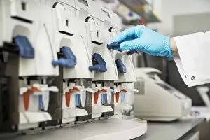
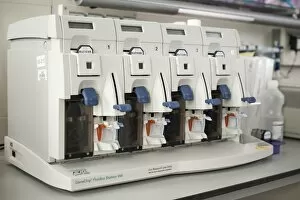
Genetic analysis. Analyst in a genetics laboratory operating a machine (fluidic station) that is being used to wash and label genetic microarrays containing samples of DNA (deoxyribonucleic acid)

Background imageImages Dated 18th December 2013: Genetic microarray preparation C019 / 0274

Genetic microarray preparation C019 / 0274
Genetic microarray preparation. Analyst pipetting a prepared DNA (deoxyribonucleic acid) sample onto a gene chip or microarray

Background imageImages Dated 18th December 2013: Bloodworm head, SEM C019 / 0229

Bloodworm head, SEM C019 / 0229
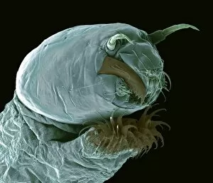
Bloodworm (Chironomus tetans) head, coloured scanning electron micrograph (SEM). The bloodworm is the freshwater larva of a midge

Background imageImages Dated 18th December 2013: Genetic analysis, pre-PCR workstation C019 / 0295

Genetic analysis, pre-PCR workstation C019 / 0295
MODEL RELEASED. Genetic analysis. Analyst in a genetics laboratory preparing samples at a pre-PCR (pre-polymerase chain reaction) workstation

Background imageImages Dated 18th December 2013: Pea moth caterpillar head, SEM C019 / 0257

Pea moth caterpillar head, SEM C019 / 0257
Pea moth caterpillar. Coloured scanning electron micrograph (SEM) of a pea moth (Cydia nigricana) caterpillar head. This caterpillar is a major pest of peas and other legumes

Background imageImages Dated 18th December 2013: Genetic analysis, pre-PCR workstation C019 / 0293

Genetic analysis, pre-PCR workstation C019 / 0293
MODEL RELEASED. Genetic analysis. Analyst in a genetics laboratory preparing samples at a pre-PCR (pre-polymerase chain reaction) workstation

Background imageImages Dated 18th December 2013: Genetic analysis, washing samples C019 / 0278

Genetic analysis, washing samples C019 / 0278
MODEL RELEASED. Genetic analysis. Analyst in a genetics laboratory operating a fluidic station that is being used to wash and label genetic microarrays containing samples of DNA

Background imageImages Dated 18th December 2013: Genetic analysis, sample preparation C019 / 0260

Genetic analysis, sample preparation C019 / 0260
Genetic analysis. Analyst in a genetics laboratory preparing blood samples before isolating DNA (deoxyribonucleic acid). This sample preparation will be done with a nucleic acid purification machine

Background imageImages Dated 18th December 2013: Genetic analysis, pre-PCR pipetting C019 / 0300

Genetic analysis, pre-PCR pipetting C019 / 0300
Genetic analysis. Automated pipetting being carried out at a pre-PCR (pre-polymerase chain reaction) workstation in a genetics laboratory

Background imageImages Dated 18th December 2013: Genetic analysis, labelling samples C019 / 0256

Genetic analysis, labelling samples C019 / 0256
MODEL RELEASED. Genetic analysis. Analyst in a genetics laboratory, labelling and scanning blood samples. DNA (deoxyribonucleic acid) will be isolated from the samples

Background imageImages Dated 18th December 2013: Genetic sample analysis C019 / 0284

Genetic sample analysis C019 / 0284
MODEL RELEASED. Genetic sample analysis. Analyst in a genetics laboratory reviewing data obtained from the analysis of a genetic microarray (held in hand)

Background imageImages Dated 18th December 2013: Genetic analysis, washing samples C019 / 0280

Genetic analysis, washing samples C019 / 0280
Genetic analysis. Machine in a genetics laboratory being used to wash and label genetic microarrays containing samples of DNA (deoxyribonucleic acid)

Background imageImages Dated 18th December 2013: Genetic karyotype analysis C019 / 0268

Genetic karyotype analysis C019 / 0268
MODEL RELEASED. Genetic karyotype analysis. Analyst in a genetics laboratory studying the results after DNA (deoxyribonucleic acid) was isolated from a sample

Background imageImages Dated 18th December 2013: Rust fungus on a bellflower leaf, SEM C019 / 0238

Rust fungus on a bellflower leaf, SEM C019 / 0238
Rust fungus. Coloured scanning electron micrograph (SEM) of a rust fungus (order Pucciniales) on a bellflower (Campanula sp.) plant

Background imageImages Dated 18th December 2013: Coccolithophore alga, SEM C019 / 0234

Coccolithophore alga, SEM C019 / 0234
Coccolithphore. Coloured scanning electron micrograph (SEM) of part of a coccolithophore unicellular marine alga. It is surrounded by a skeleton (coccosphere) of calcium carbonate plates (coccoliths)

Background imageImages Dated 18th December 2013: Genetic microarray preparation C019 / 0273

Genetic microarray preparation C019 / 0273
MODEL RELEASED. Genetic microarray preparation. Analyst pipetting a prepared DNA (deoxyribonucleic acid) sample onto a gene chip or microarray

Background imageImages Dated 18th December 2013: Genetic microarray preparation C019 / 0272

Genetic microarray preparation C019 / 0272
MODEL RELEASED. Genetic microarray preparation. Analyst pipetting a prepared DNA (deoxyribonucleic acid) sample onto a gene chip or microarray

Background imageImages Dated 18th December 2013: Climacostomum protozoan, SEM C019 / 0243

Climacostomum protozoan, SEM C019 / 0243
Climacostomum sp. ciliate protozoan, coloured scanning electron micrograph (SEM). These tiny single-celled organisms are found in freshwater and marine habitats

Background imageImages Dated 18th December 2013: Microglial white blood cell, SEM C019 / 0245

Microglial white blood cell, SEM C019 / 0245
Microglia. Coloured scanning electron micrograph (SEM) of an activated microglial white blood cell. This immune system cell is found in the brain and spinal cord

Background imageImages Dated 18th December 2013: Genetic metaphase analysis C019 / 0288

Genetic metaphase analysis C019 / 0288
Genetic metaphase analysis. Equipment used in a genetics laboratory to analyse the metaphase stage of cell division. During this stage

Background imageImages Dated 18th December 2013: Genetic analysis, DNA isolation C019 / 0267

Genetic analysis, DNA isolation C019 / 0267
Genetic analysis of blood. Automated nucleic acid purification machine being used to purify and isolate DNA (deoxyribonucleic acid) from blood samples

Background imageImages Dated 18th December 2013: Gastrotrich, SEM C019 / 0232

Gastrotrich, SEM C019 / 0232
Gastrotrich. Coloured scanning electron micrograph (SEM) of a Chaetonotus sp. gastrotrich. Gastrotrichs are microscopic, worm-like animals found in both freshwater and marine habitats

Background imageImages Dated 18th December 2013: Genetic microarray, analysis machine C019 / 0282

Genetic microarray, analysis machine C019 / 0282
Genetic microarray, analysis machine. Analyst placing a prepared gene chip or microarray into an analysis machine. Samples of DNA (deoxyribonucleic acid) have been added to the microarray

Background imageImages Dated 18th December 2013: Genetic analysis, DNA isolation C019 / 0265

Genetic analysis, DNA isolation C019 / 0265
MODEL RELEASED. Genetic analysis. Analyst using a nucleic acid purification machine to purify and isolate DNA (deoxyribonucleic acid) from blood samples

Background imageImages Dated 18th December 2013: Coccolithophore alga, SEM C019 / 0224

Coccolithophore alga, SEM C019 / 0224
Coccolithphore. Coloured scanning electron micrograph (SEM) of a coccolithophore unicellular marine alga. It is surrounded by a skeleton (coccosphere) of calcium carbonate plates (coccoliths)

Background imageImages Dated 18th December 2013: Genetic microarray, analysis machine C019 / 0281

Genetic microarray, analysis machine C019 / 0281
MODEL RELEASED. Genetic microarray, analysis machine. Analyst placing a prepared gene chip or microarray into an analysis machine

Background imageImages Dated 18th December 2013: 2013 Kia Sportage

2013 Kia Sportage

Background imageImages Dated 18th December 2013: 2013 Kia Sportage

2013 Kia Sportage dashboard

Background imageImages Dated 18th December 2013: 2013 Kia Sportage

2013 Kia Sportage

Background imageImages Dated 18th December 2013: 2013 Kia Sportage

2013 Kia Sportage

Background imageImages Dated 18th December 2013: 2013 Kia Sportage

2013 Kia Sportage

Background imageImages Dated 18th December 2013: B&H Cup Final: Lancs bt Kent 35r

B&H Cup Final: Lancs bt Kent 35r
Cricket - 1995 Benson & Hedges Cup Final - Lancashire bt Kent by 35 runs Lancashire captain Mike Watkinson with the trophy, with Ian Austin, left, John Crawley, behind, and Mike Atherton, right


All Professionally Made to Order for Quick Shipping


-

Why Choose Us?

We are a leading provider of Art Prints and Photo Gifts since 2003, working in partnership with a range of Sporting Clubs, Charities, Museums and Picture Libraries. A large share of profits from any of their images will go directly towards supporting that charity or club. Our archive of images is carefully curated to bring you a wide range of subjects, including landscapes, wildlife, architecture, and more. We ship from our partner labs in the UK, USA, EU (Netherlands) and Australia.
+

How do I place an order?

Ordering is quick & easy - Just follow 5 Simple Steps:
  1. Find your image: Use our search box or browse our online photo Collections to find the image you want.

  2. Choose your print format: Select your desired product and add it to your cart.

  3. Enter your details: If you're a returning customer, simply enter your email address and password, and we'll fill in your billing and shipping address details. All personal details are held securely and are fully GDPR compliant. As standard, we remove all Personally Identifiable Information after 12 months.

  4. Pay for your purchase: We use state-of-the-art security for online shopping and do not have access to your card details.

  5. Sit back and relax: We'll email you confirmation of your order and when it's dispatched. Registered customers can also track orders in the 'My Account' area.

+

How do I pay for an order?

You can pay for your order with most of the major credit and debit cards, or PayPal. For added security, major financial institutions process payment details separately and securely on our behalf. We do not have access to your online payment card details. Online payments are preferred however we do take phone orders. For UK customers only we accept cheques issued against a UK bank.
+

Is my data safe?

We take data security very seriously. We do not have access to your full card details and all payments are requested over a fully secure connection. Additionally, we fully comply with current European and GDPR legislation, and automatically remove all personal data after at most 12 months (unless you have an account with us and have signed in recently). We also have a strict opt-in policy and would never sell your personal details. Your data is only used to fulfill your order promptly and efficiently.
+

Is wall art ready to hang?

For quick and easy installation all wall art, including framed prints, canvas prints and metal prints are supplied with a ready to hang solution on the back. Generally, saw tooth hangers are applied as they allow wall art to hang flush against the wall. The serrated edge of the hanger prevents the frame from shifting or tilting when hung.
+

Are the photo prints fade resistant?

Yes, we use archival quality photo paper photographic paper for vivid reproduction Prints are an accurate representations of the original artwork, which is preserved for artistic character and authenticity. We guarantee they match previews shown on our web site