Fraud Blocker Skip to main content

Home > Science > SEM

SEM Collection (page 6)

7,669 items

Background imageSEM Collection: Eye muscle, SEM

Eye muscle, SEM
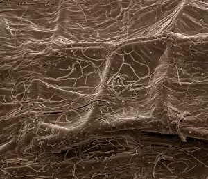
Eye muscle, coloured scanning electron micrograph (SEM). This is one of the rectus muscles of the eye. These muscles control the movement of the eyeball within the orbit (the eye socket)

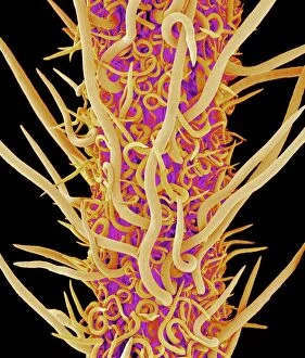
Background imageSEM Collection: Banksia stem, SEM

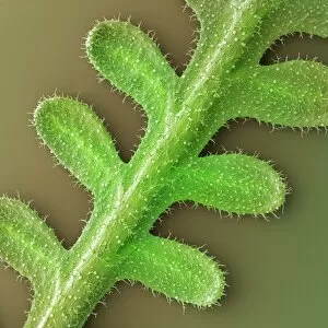
Banksia stem, SEM
Banksia stem, coloured scanning electron micrograph (SEM). The stem is covered in fine hairs

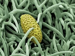
Background imageSEM Collection: Gorse stigma with pollen grains, SEM

Gorse stigma with pollen grains, SEM
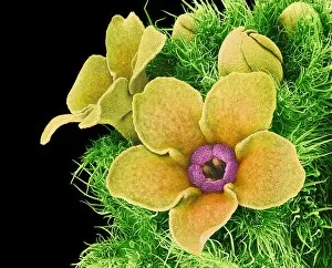
Gorse (Ulex europaeus) stigma (green) with pollen grains (yellow), coloured scanning electron micrograph (SEM). Pollen grains are reproductive structures produced by the male parts of flowering

Background imageSEM Collection: Lily pollen grain on rosemary leaf, SEM

Lily pollen grain on rosemary leaf, SEM
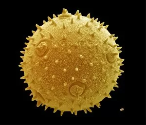
Lily pollen grain on rosemary leaf. Coloured scanning electron micrograph (SEM) of a pollen grain (yellow) from a lily flower (Lilium sp.)

Background imageSEM Collection: Gorse flower bud, SEM

Gorse flower bud, SEM
Gorse (Ulex europaeus) flower bud, coloured scanning electron micrograph (SEM). One of the petals (yellow) is seen enclosed in the flower casing (calyx)

Background imageSEM Collection: Forsythia pollen grains, SEM

Forsythia pollen grains, SEM
Forsythia pollen grains arranged in a spiral, coloured scanning electron micrograph (SEM). Pollen grains are reproductive structures produced by the male parts of flowering plants

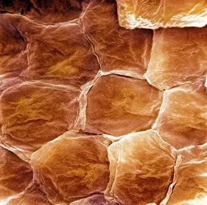
Background imageSEM Collection: Skin surface, SEM

Skin surface, SEM
Skin surface. Coloured scanning electron micrograph (SEM) of squamous cells on the surface of the skin. These are flat, keratinised

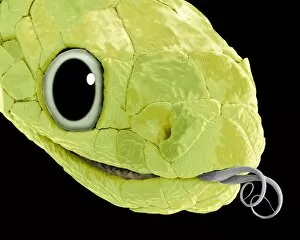
Background imageSEM Collection: Snake head, SEM

Snake head, SEM
Snake (suborder Serpentes) head, coloured scanning electron micrograph (SEM). The snakes forked tongue (right) is used to detect prey by picking up scent particles from the air

Background imageSEM Collection: Chickweed pollen grains, SEM

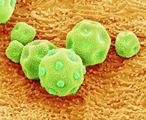
Chickweed pollen grains, SEM
Chickweed pollen grains. Coloured scanning electron micrograph (SEM) of pollen grains (green) from a chickweed flower (Stellaria media)

Background imageSEM Collection: Flame lily pollen, SEM

Flame lily pollen, SEM
Flame lily (Gloriosa sp.) pollen grains, coloured scanning electron micrograph (SEM). The grains have a highly sculptured outer wall (exine)

Background imageSEM Collection: Mixture of flower and grass seeds, SEM

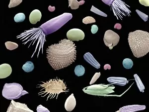
Mixture of flower and grass seeds, SEM
Mixture of flower and grass seeds, coloured scanning electron micrograph (SEM). This is a mixture of seeds for wild meadow plants. It includes flower and grass seeds

Background imageSEM Collection: Hellebore pollen, SEM

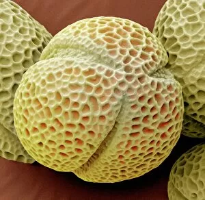
Hellebore pollen, SEM
Hellebore (Helleborus sp.) pollen grains, coloured scanning electron micrograph (SEM). Magnification: x2000 when printed at 10 centimetres wide

Background imageSEM Collection: Calcium phosphate crystal, SEM

Calcium phosphate crystal, SEM
Calcium phosphate crystal, coloured scanning electron micrograph (SEM). Crystalline materials have their atoms placed in regular lattices that can form geometric shapes like this

Background imageSEM Collection: E. coli 0157: H7 bacteria

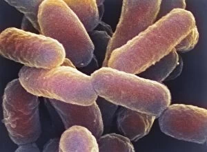
E. coli 0157: H7 bacteria
E. coli 0157:H7 bacteria. Coloured scanning electron micrograph (SEM) of Escherichia coli 0157:H7 bacteria, cause of foodborne illness

Background imageSEM Collection: Salmonella bacterium dividing, SEM

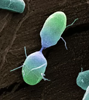
Salmonella bacterium dividing, SEM
Salmonella bacterium dividing. Coloured scanning electron micrograph (SEM) of of a Salmonella bacterium dividing. The two new daughter cells are seen at upper right and lower left

Background imageSEM Collection: Bacteria infecting a macrophage, SEM

Bacteria infecting a macrophage, SEM
Bacteria infecting a macrophage. Coloured scanning electron micrograph (SEM) of Mycobacterium tuberculosis bacteria (purple) infecting a macrophage white blood cell

Background imageSEM Collection: Common horsetail spore, SEM

Common horsetail spore, SEM
Common horsetail spore. Coloured scanning electron micrograph (SEM) of a spore of a common horsetail (Equisetum arvense) plant

Background imageSEM Collection: Spores of field horsetail

Spores of field horsetail
False colour scanning electron micrograph of spores of the field horsetail, Equisetum arvense. The horsetail is the only living type of a primitive family of plants, the Sphenopsids

Background imageSEM Collection: Xylem tissue, SEM

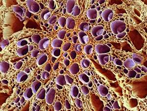
Xylem tissue, SEM
Xylem tissue. Coloured scanning electron micrograph (SEM) of a section through xylem tissue from a dicotyledon rootlet. Xylem vessels (purple)

Background imageSEM Collection: Root of a germinating red chard seed, SEM

Root of a germinating red chard seed, SEM
Root of a germinating red chard seed. Coloured scanning electron micrograph (SEM) of a red chard seed (Beta vulgaris var. cicla) root

Background imageSEM Collection: Lily petal, SEM

Lily petal, SEM
Lily petal. Coloured scanning electron micrograph (SEM) of the surface of a lily (family Liliaceae). Magnification x58 at 6x7cm size

Background imageSEM Collection: Pollen tubes of lily pollen, SEM

Pollen tubes of lily pollen, SEM
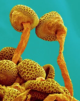
Pollen tubes of lily pollen, coloured scanning electron micrograph (SEM). Two pollen grains (brown) have sprouted pollen tubes (orange)

Background imageSEM Collection: Germinating lily pollen, SEM

Germinating lily pollen, SEM
Germinating lily pollen. Coloured scanning electron micrograph (SEM) of lily (Lilium sp.) pollen grains that have sprouted pollen tubes. Pollen contains the male sex cells of a plant

Background imageSEM Collection: Flower, SEM

Flower, SEM
Flower. Coloured scanning electron micrograph (SEM) of a flower

Background imageSEM Collection: Spider lily flower stamen, SEM

Spider lily flower stamen, SEM
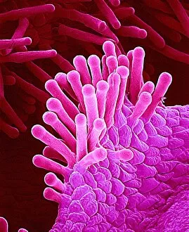
Spider lily flower stamen. Coloured scanning electron micrograph (SEM) of a cilia (hair-like structures) from a spider lily flower (Hymenocallis sp.) stamen

Background imageSEM Collection: Columbine flower stamens, SEM

Columbine flower stamens, SEM
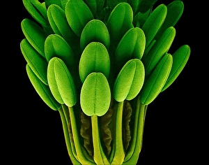
Columbine flower stamens. Coloured scanning electron micrograph (SEM) of the stamens (male sexual parts) of a columbine flower (Aquilegia sp.)

Background imageSEM Collection: Red Clover pollen

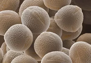
Red Clover pollen. Coloured scanning electron micrograph (SEM) of pollen grains of Red Clover, Trifolium pratense. The outer wall (exine) of each grain has a netted ornamentation

Background imageSEM Collection: Leaf oil glands, SEM

Leaf oil glands, SEM
Leaf oil glands. Coloured scanning electron micrograph (SEM) of a thyme plants leaf surface (Thymus vulgaris). Two types of leaf trichomes (hairs) are seen

Background imageSEM Collection: French lavender leaf, SEM

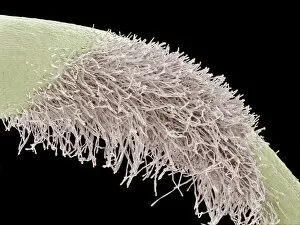
French lavender leaf, SEM
French lavender (Lavandula dentata) leaf. Coloured scanning electron micrograph (SEM) of a section of a French lavender leaf, showing the tooth-like structures (trichomes)

Background imageSEM Collection: False-colour SEM of chickweed flower

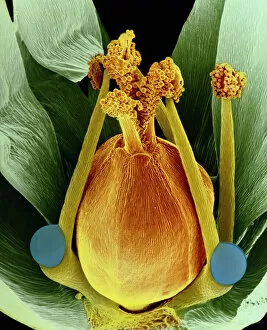
False-colour SEM of chickweed flower
False-colour scanning electron micrograph of the reproductive structures of a flower of the chickweed, Stellaria media. The bulbous carpel, with the short-stalked female stigmas emerging from it

Background imageSEM Collection: Grape tendril, SEM

Grape tendril, SEM
Grape tendril. Coloured scanning electron micrograph (SEM) of a curled grape (Vitis sp.) tendril. Tendrils are specialised thread-like stems or leaves that are used by climbing plants for support

Background imageSEM Collection: Yarrow flower petal, SEM

Yarrow flower petal, SEM
Yarrow flower petal. Coloured scanning electron micrograph (SEM) of a petal from the flower of a Japanese type of yarrow (Achillea alpina)

Background imageSEM Collection: Daisy bud, SEM

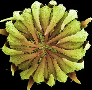
Daisy bud, SEM
Daisy. Coloured scanning electron micrograph of a daisy bud (family Asteraceae, formerly Compositae). The protective bracts (green) cover clusters of tiny flowers, or florets

Background imageSEM Collection: Zinnia flower petal, SEM

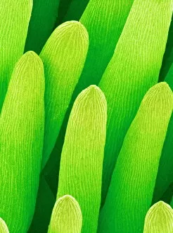
Zinnia flower petal, SEM
Zinnia flower petal. Coloured scanning electron micrograph (SEM) of a Zinnia sp. petal. The projections on the petal help to prevent water loss

Background imageSEM Collection: Pollination, SEM

Pollination, SEM
Pollination. Coloured scanning electron micrograph (SEM) of part of a flowers pistil with pollen grains attached. The pistil is the female reproductive part of a flower

Background imageSEM Collection: French lavender leaf pore, SEM

French lavender leaf pore, SEM
French lavender leaf pore. Coloured scanning electron micrograph (SEM) of an open stoma (centre, black). Stomata are pores that open and close in order to regulate gas exchange in a plant

Background imageSEM Collection: Periwinkle petal surface, SEM

Periwinkle petal surface, SEM
Periwinkle petal. Coloured scanning electron micrograph (SEM) of the surface of a periwinkle flower petal (family Apocynaceae)

Background imageSEM Collection: Wild flower, SEM

Wild flower, SEM
Wild flower. Coloured scanning electron micrograph (SEM) of an unidentified flower. Magnification: x38 at 6x7cm size

Background imageSEM Collection: Marrow and forget-me-not pollen, SEM

Marrow and forget-me-not pollen, SEM
Pollen grains. Coloured scanning electron micrograph (SEM) of a pollen grain from a marrow plant (Cucurbita sp. left) and a forget-me-not plant (Myosotis sp. lower right)

Background imageSEM Collection: Diatom alga, SEM

Diatom alga, SEM
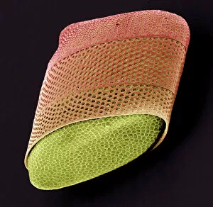
Diatom. Coloured scanning electron micrograph (SEM) of a Biddulphia sp. diatom. This is a marine planktonic unicellular alga. It has a mineralised cell wall (frustule) divided into two halves

Background imageSEM Collection: False colour SEM of moss leaves; green

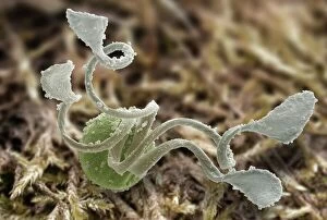
False colour SEM of moss leaves; green
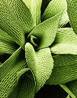
False-colour scanning electron micrograph of a vegetative shoot of the moss Physcomitrella patens. The leaves of mosses are very simple compared with those of higher plants

Background imageSEM Collection: False col. SEM of crocidolite fibres

False col. SEM of crocidolite fibres
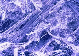
False-colour scanning electron micrograph of fibres of blue asbestos, or crocidolite. All asbestos varieties are silicates (amphibole minerals), the most common being crocidolite

Background imageSEM Collection: Silver crystals

Silver crystals. Coloured scanning electron micrograph (SEM) of crystals of silver (symbol Ag, atomic number 47). Silver is a lustrous, precious metal which has been known since pre-history

Background imageSEM Collection: Scanning Electron Micrograph (SEM): Mycorrhiza fungus in plant root; Magnification x 700

Scanning Electron Micrograph (SEM): Mycorrhiza fungus in plant root; Magnification x 700 (if printed A4, 29. 7 cm wide)
LRDS-472 Scanning Electron Micrograph (SEM): Mycorrhiza fungus in plant root; Magnification x 700 (if printed A4, 29.7 cm wide) David Spears (Last Refuge)

Background imageSEM Collection: Scanning Electron micrograph (SEM): Leaf Hairs - of dicot plant showing gutation water droplets;

Scanning Electron micrograph (SEM): Leaf Hairs - of dicot plant showing gutation water droplets; Magnification unknown
LRDS-462 Scanning Electron micrograph (SEM): Leaf Hairs - of dicot plant showing gutation water droplets Magnification unknown David Spears (Last Refuge)

Background imageSEM Collection: Scanning Electron Micrograph (SEM): Driver Ant - Magnification x 100 (if print A4 size: 29)

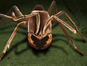
Scanning Electron Micrograph (SEM): Driver Ant - Magnification x 100 (if print A4 size: 29. 7 cm wide)
LRDS-409 Scanning Electron Micrograph (SEM): Driver Ant Magnification x 100 (if print A4 size: 29.7 cm wide) Dorylus sp. David Spears (Last Refuge)

Background imageSEM Collection: Scanning Electron Micrograph (SEM): Powderpost Beetle - Magnification x 50 (if print A4 size: 29)

Scanning Electron Micrograph (SEM): Powderpost Beetle - Magnification x 50 (if print A4 size: 29. 7 cm wide)
LRDS-418 Scanning Electron Micrograph (SEM): Powderpost Beetle Magnification x 50 (if print A4 size: 29.7 cm wide) Lyetus brunneus David Spears (Last Refuge)

Background imageSEM Collection: Scanning Electron Micrograph (SEM): Mosquito, Female; Magnification x 55 (A4 size: 29. 7 cm width)

Scanning Electron Micrograph (SEM): Mosquito, Female; Magnification x 55 (A4 size: 29. 7 cm width)
LRDS-83 Mosquito, Female Scanning Electron Micrograph (SEM) Anopheles sp. Magnification x 55 (A4 size: 29.7 cm width) Coloured by hand to enhance natural features


All Professionally Made to Order for Quick Shipping


-

Why Choose Us?

We are a leading provider of Art Prints and Photo Gifts since 2004, working in partnership with a range of Sporting Clubs, Charities, Museums and Picture Libraries. A large share of profits from any of their images will go directly towards supporting that charity or club. Our archive of images is carefully curated to bring you a wide range of subjects, including landscapes, wildlife, architecture, and more. We ship from our partner labs in the UK, USA, EU (Netherlands) and Australia.
+

How do I place an order?

Ordering is quick & easy - Just follow 5 Simple Steps:
  1. Find your image: Use our search box or browse our online photo Collections to find the image you want.

  2. Choose your print format: Select your desired product and add it to your cart.

  3. Enter your details: If you're a returning customer, simply enter your email address and password, and we'll fill in your billing and shipping address details. All personal details are held securely and are fully GDPR compliant. As standard, we remove all Personally Identifiable Information after 12 months.

  4. Pay for your purchase: We use state-of-the-art security for online shopping and do not have access to your card details.

  5. Sit back and relax: We'll email you confirmation of your order and when it's dispatched. Registered customers can also track orders in the 'My Account' area.

+

How do I pay for an order?

You can pay for your order with most of the major credit and debit cards, or PayPal. For added security, major financial institutions process payment details separately and securely on our behalf. We do not have access to your online payment card details. Online payments are preferred however we do take phone orders. For UK customers only we accept cheques issued against a UK bank.
+

Is my data safe?

We take data security very seriously. We do not have access to your full card details and all payments are requested over a fully secure connection. Additionally, we fully comply with current European and GDPR legislation, and automatically remove all personal data after at most 12 months (unless you have an account with us and have signed in recently). We also have a strict opt-in policy and would never sell your personal details. Your data is only used to fulfill your order promptly and efficiently.
+

Is wall art ready to hang?

For quick and easy installation all wall art, including framed prints, canvas prints and metal prints are supplied with a ready to hang solution on the back. Generally, saw tooth hangers are applied as they allow wall art to hang flush against the wall. The serrated edge of the hanger prevents the frame from shifting or tilting when hung.
+

Are the photo prints fade resistant?

Yes, we use archival quality photo paper photographic paper for vivid reproduction Prints are an accurate representations of the original artwork, which is preserved for artistic character and authenticity. We guarantee they match previews shown on our web site