Fraud Blocker Skip to main content

Home > Science > SEM

SEM Collection (page 3)

1,336 items

Background imageSEM Collection: Picture No. 11675612

Picture No. 11675612
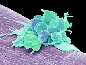
Scanning Electron Micrograph (SEM): Human White and Red Blood Cells. Date

Background imageSEM Collection: Picture No. 11675599

Picture No. 11675599
Dark Field Light Micrograph: Lancet Liver Fluke. Date

Background imageSEM Collection: Picture No. 10876997

Picture No. 10876997
Scanning Electron Micrograph (SEM): Human Skin with Hair Follicles Date

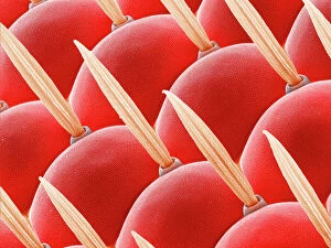
Background imageSEM Collection: Compound eye of a fly, SEM Z340 / 0698

Compound eye of a fly, SEM Z340 / 0698
Compound eye. Coloured scanning electron micrograph (SEM) of the surface of a compound eye from a fruit fly (Drosophila busckii). The eye consists of many rounded lenses known as ommatidia

Background imageSEM Collection: Moss capsule (Homalothecium sericeum) SEM

Moss capsule (Homalothecium sericeum) SEM
Moss spore capsule. Coloured scanning electron micrograph (SEM) of part of the open mouth of a capsule (spore case) of a the moss Homalothecium sericeum

Background imageSEM Collection: Red and white blood cells, SEM

Red and white blood cells, SEM
Human red and white blood cells, coloured scanning electron micrograph (SEM). Magnification x5167 at an image size of 10 cm wide

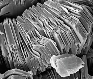
Background imageSEM Collection: Kaolinite

Kaolinite
Scanning electron microscope image of kaolinite (x 4.00K). Its a common phyllosilicate mineral, its structure is composed of silicate sheets bonded to aluminum oxide/hydroxide layers

Background imageSEM Collection: Sarcoptes scabiei, scabies mite

Sarcoptes scabiei, scabies mite
Scanning electron microscope image of an itch or scabies mite, a parasite that infests a wide variety of mammalian hosts including humans

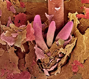
Background imageSEM Collection: Eyelash mite tails, SEM

Eyelash mite tails, SEM
Eyelash mite tails. Coloured scanning electron micrograph (SEM) of the tails (pink) of three eyelash, or follicle, mites (Demodex folliculorum), protruding from a human hair follicle

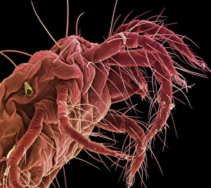
Background imageSEM Collection: Spider mite, SEM

Spider mite, SEM
Spider mite. Coloured scanning electron micrograph (SEM) of a spider mite (Tetranychus sp.). It is an economic pest and plant parasite, found on vegetable crops, fruit trees and flowers

Background imageSEM Collection: Moth antenna, SEM

Moth antenna, SEM
Moth antenna. Coloured scanning electron micro- graph (SEM) of an unidentified moths antenna. It is covered in tiny sensory hairs and sense organs that sense movement and smell

Background imageSEM Collection: SEM of section through human skin

SEM of section through human skin
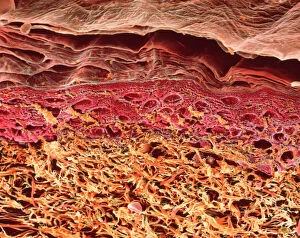
Skin. Coloured scanning electron micrograph of a section through the human skin. The uppermost layers of the skin (pink and red) make up the epidermis

Background imageSEM Collection: Tendon, SEM

Tendon, SEM
Tendon, coloured scanning electron micrograph (SEM), showing bundles of collagen fibres. Magnification: x5000 when printed at 10 centimetres wide

Background imageSEM Collection: Macrophage engulfing TB bacteria, SEM

Macrophage engulfing TB bacteria, SEM
Macrophage engulfing TB bacteria. Coloured scanning electron micrograph (SEM) of a macrophage white blood cell (purple) engulfing a tuberculosis (Mycobacterium tuberculosis) bacterium (pink)

Background imageSEM Collection: Scanning Electron Micrograph (SEM): Crab Spider, Magnification x 140 (A4 size: 29. 7 cm width)

Scanning Electron Micrograph (SEM): Crab Spider, Magnification x 140 (A4 size: 29. 7 cm width)
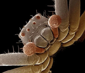
LRDS-120 Crab Spider Scanning Electron Micrograph (SEM) Xysticus sp. Magnification x 140 (A4 size: 29.7 cm width) Coloured by hand to enhance natural features

Background imageSEM Collection: Scanning Electron Micrograph (SEM): House Fly, Magnification x 40 (A4 size: 29. 7 cm width)

Scanning Electron Micrograph (SEM): House Fly, Magnification x 40 (A4 size: 29. 7 cm width)
LRDS-72 House Fly Scanning Electron Micrograph (SEM) Musca domestica Magnification x 40 (A4 size: 29.7 cm width) Coloured by hand to enhance natural features

Background imageSEM Collection: Diatom detail, SEM

Diatom detail, SEM
Diatom. Coloured scanning electron micrograph (SEM) of detail of the silica wall of a diatom. Diatoms may be extremely abundant in both freshwater

Background imageSEM Collection: Activated platelets, SEM

Activated platelets, SEM
Activated platelets. Coloured scanning electron micrograph (SEM) of activated platelets attached to surgical gauze. Platelets are tiny blood cells that help the body form clots to stop bleeding

Background imageSEM Collection: Red flour beetle in flight

Red flour beetle in flight
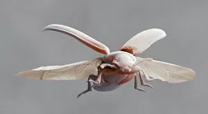
Red flour beetle. Coloured scanning electron micrograph of a red flour beetle (Tribolium castaneum) in flight. One of the most significant pests of dried foods and the milling industry

Background imageSEM Collection: Activated macrophage, SEM C015 / 6375

Activated macrophage, SEM C015 / 6375
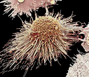
Activated macrophage. Coloured scanning electron micrograph (SEM) of an active macrophage white blood cell. Macrophages are cells of the bodys immune system that are found in the tissues rather than

Background imageSEM Collection: Seller of oysters from the store Prunier. Chromolithography signed SEM (1863 - 1934)

Seller of oysters from the store Prunier. Chromolithography signed SEM (1863 - 1934)
LSE4112452 Seller of oysters from the store Prunier. Chromolithography signed SEM (1863 - 1934). by Sem (Georges Goursat) (1863-1934); Private Collection; (add.info)

Background imageSEM Collection: Microfossils, SEM

Microfossils, SEM
Sand microfossils. Coloured scanning electron micrograph (SEM) of microfossils from maldives beach sand. Microfossils are roughly 0.05 to 2mm in size

Background imageSEM Collection: Picture No. 11675582

Picture No. 11675582
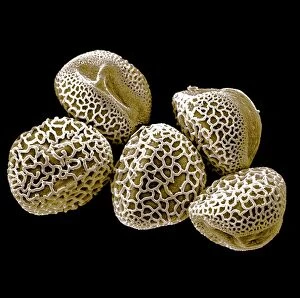
Scanning Electron micrograph (SEM): Lily Pollen grains. Date

Background imageSEM Collection: Picture No. 11675585

Picture No. 11675585
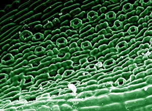
Scanning Electron micrograph (SEM)showing stomata on a Yew Leaf. Date

Background imageSEM Collection: Adam and Eves Family Tree, 1556 (engraving)

Adam and Eves Family Tree, 1556 (engraving)
XJF476254 Adam and Eves Family Tree, 1556 (engraving) by English School, (16th century); Private Collection; English, out of copyright

Background imageSEM Collection: Nerve cells, SEM

Nerve cells, SEM
Nerve cells. Coloured scanning electron micrograph (SEM) of nerve cells, known as neurones. Nerve cells occur in the brain, spinal cord, and in ganglia

Background imageSEM Collection: Picture No. 10873704

Picture No. 10873704
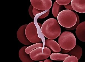
Sleeping Sickness Parasite in red blood cells (Trypanosoma sp. ) Date

Background imageSEM Collection: Blood clot, SEM C016 / 9747

Blood clot, SEM C016 / 9747
Blood clot, coloured scanning electron micrograph (SEM). Red blood cells (erythrocytes) are trapped within a fibrin protein mesh (beige)

Background imageSEM Collection: Tropical ants, SEM

Tropical ants, SEM
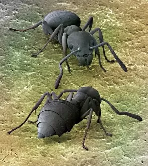
Tropical ants. Coloured scanning electron micrograph (SEM) of two small, tropical ants (family Formicidae, 2 millimetres long)

Background imageSEM Collection: Fruit fly, SEM Z340 / 0699

Fruit fly, SEM Z340 / 0699
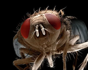
Fruit fly. Coloured scanning electron micrograph (SEM) of the head of a fruit fly (Drosophila busckii). Its two compound eyes (red) are seen on either side of the head

Background imageSEM Collection: Dust mite

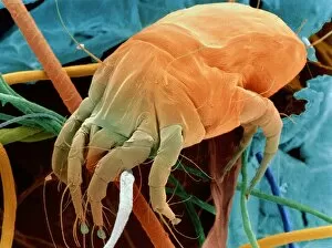
Dust mite. Coloured Scanning Electron Micrograph (SEM) of a dust mite Dermatophagoides sp. in a sample of household dust. It is crawling among hair, fibres and other dust debris

Background imageSEM Collection: Stalk-eyed fly, SEM C014 / 4882

Stalk-eyed fly, SEM C014 / 4882
Stalk-eyed fly. Coloured scanning electron micrograph (SEM) of the head of a stalk-eyed fly (family Diopsidae). Stalk-eyed flies are mostly tropical insects

Background imageSEM Collection: Rod and cone cells of the eye, SEM C014 / 4866

Rod and cone cells of the eye, SEM C014 / 4866
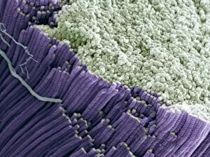
Rod and cone cells of the eye. Coloured scanning electron micrograph (SEM) of rod and cone cells in the retina of a mammalian eye

Background imageSEM Collection: Baking soda crystals, SEM C016 / 8041

Baking soda crystals, SEM C016 / 8041
Sodium bicarbonate. Coloured scanning electron micrograph (SEM) of crystals of sodium bicarbonate (or sodium hydrogen carbonate). Sodium bicarbonate is a white solid commonly used in baking powder

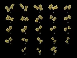
Background imageSEM Collection: Full set of male chromosomes, SEM

Full set of male chromosomes, SEM
Set of human chromosomes, coloured scanning election micrograph (SEM). Chromosomes are a packaged form of the genetic material deoxyribonucleic acid (DNA)

Background imageSEM Collection: Foraminifera, SEM

Foraminifera, SEM
Foraminifera. Coloured scanning electron micrograph (SEM) of a selection of foraminiferans, including Oolina hexagona (bottom right) and a number of Lagena sp

Background imageSEM Collection: English oak leaf pores, SEM

English oak leaf pores, SEM
English oak leaf pores. Coloured scanning electron micrograph (SEM) of stomata (round) on the underside of a leaf from an English oak (Quercus robur) tree

Background imageSEM Collection: Thyme leaf oil gland

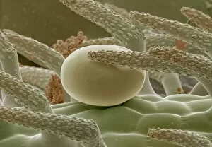
Thyme leaf oil gland. Coloured scanning electron micrograph (SEM) of part of the leaf surface of the herb thyme, Thymus vulgaris, showing an oil gland

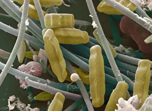
Background imageSEM Collection: SEM of diatoms and blue-green algae

SEM of diatoms and blue-green algae
Diatoms & blue-green algae. Coloured scanning electron micrograph (SEM) of groups of Navicula sp. diatoms (brown). Also seen in the image are strands of blue-green algae (blue)

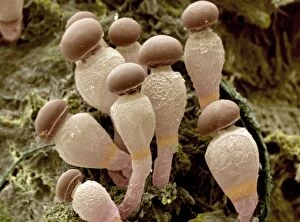
Background imageSEM Collection: Pilobolus fungus

Pilobolus fungus
Fungus sporangiophores. Coloured scanning electron micrograph (SEM) of sporangiophores of a Pilobolus sp. fungus, growing on dung

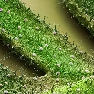
Background imageSEM Collection: French lavender leaf surface, SEM

French lavender leaf surface, SEM
French lavender leaf surface. Coloured scanning electron micrograph (SEM) of a section of French lavender (Lavandula dentata) leaf, showing the tooth-like structures (trichomes)

Background imageSEM Collection: Acanthoica acanthifera, coccosphere

Acanthoica acanthifera, coccosphere
Scanning electron microscope (SEM) image of a coccosphere, collected in the North Atlantic (x 25, 000 on negative). Artificially coloured by computer

Background imageSEM Collection: Human red blood corpuscles

Human red blood corpuscles
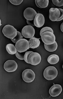
Scanning electron microscope (SEM) of red blood cells showing their characteristic biconcave shape which increases the surface area for diffusion

Background imageSEM Collection: Globorotalia scitula, foraminifera fossil

Globorotalia scitula, foraminifera fossil
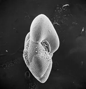
Scanning electron microscope (SEM) image showing a fossilised planktonic species of foraminifera

Background imageSEM Collection: Scyliorhinus canicula, dogfish

Scyliorhinus canicula, dogfish
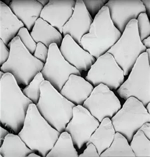
Scanning electron microscope (SEM) image of the scales of a dogfish (x 40)

Background imageSEM Collection: Needle playing a record, SEM

Needle playing a record, SEM
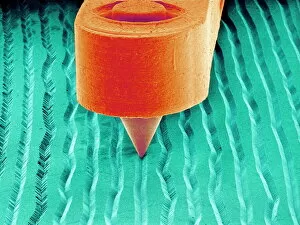
Needle playing a record. Coloured scanning electron micrograph (SEM) of the needle (stylus) of a record player in a groove on a record. A record is used to store sound

Background imageSEM Collection: Ragworm mouth, SEM

Ragworm mouth, SEM
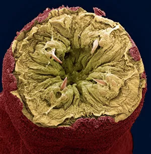
Ragworm mouth. Coloured scanning electron micrograph (SEM) of the mouth of a ragworm (Nereis sp.). Four teeth (cream) are seen around the opening. These worms are used commercially as fishing bait

Background imageSEM Collection: Nematode worm, SEM

Nematode worm, SEM
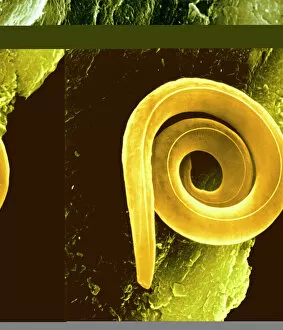
Nematode worm, coloured scanning electron micrograph (SEM). Nematode worms are small worms that are unsegmented. Depending on the species, a nematode worm may be parasitic or free-living


All Professionally Made to Order for Quick Shipping


-

Why Choose Us?

We are a leading provider of Art Prints and Photo Gifts since 2003, working in partnership with a range of Sporting Clubs, Charities, Museums and Picture Libraries. A large share of profits from any of their images will go directly towards supporting that charity or club. Our archive of images is carefully curated to bring you a wide range of subjects, including landscapes, wildlife, architecture, and more. We ship from our partner labs in the UK, USA, EU (Netherlands) and Australia.
+

How do I place an order?

Ordering is quick & easy - Just follow 5 Simple Steps:
  1. Find your image: Use our search box or browse our online photo Collections to find the image you want.

  2. Choose your print format: Select your desired product and add it to your cart.

  3. Enter your details: If you're a returning customer, simply enter your email address and password, and we'll fill in your billing and shipping address details. All personal details are held securely and are fully GDPR compliant. As standard, we remove all Personally Identifiable Information after 12 months.

  4. Pay for your purchase: We use state-of-the-art security for online shopping and do not have access to your card details.

  5. Sit back and relax: We'll email you confirmation of your order and when it's dispatched. Registered customers can also track orders in the 'My Account' area.

+

How do I pay for an order?

You can pay for your order with most of the major credit and debit cards, or PayPal. For added security, major financial institutions process payment details separately and securely on our behalf. We do not have access to your online payment card details. Online payments are preferred however we do take phone orders. For UK customers only we accept cheques issued against a UK bank.
+

Is my data safe?

We take data security very seriously. We do not have access to your full card details and all payments are requested over a fully secure connection. Additionally, we fully comply with current European and GDPR legislation, and automatically remove all personal data after at most 12 months (unless you have an account with us and have signed in recently). We also have a strict opt-in policy and would never sell your personal details. Your data is only used to fulfill your order promptly and efficiently.
+

Is wall art ready to hang?

For quick and easy installation all wall art, including framed prints, canvas prints and metal prints are supplied with a ready to hang solution on the back. Generally, saw tooth hangers are applied as they allow wall art to hang flush against the wall. The serrated edge of the hanger prevents the frame from shifting or tilting when hung.
+

Are the photo prints fade resistant?

Yes, we use archival quality photo paper photographic paper for vivid reproduction Prints are an accurate representations of the original artwork, which is preserved for artistic character and authenticity. We guarantee they match previews shown on our web site