Home > Popular Themes > Human Body
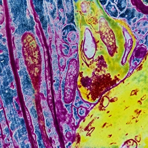
Synapse nerve junction, TEM

![]()
Wall Art and Photo Gifts from Science Photo Library
Synapse nerve junction, TEM
Synapse. Coloured transmission electron micrograph (TEM) of a synapse, a junction between two nerve cells, in the brain. At a synapse an electrical signal is transmitted from one cell to the next in only one direction. The nerve cells are green, with the pre-synaptic cell at lower right and the post-synaptic cell at upper left. Mitochondria, supplying the cells with energy, are purple. When an electrical signal reaches a synapse it triggers the release of neurotransmitter chemicals from vesicles (red) at the end of the presynaptic cell. The neurotransmitters cross a microscopic gap and bind to receptors on the post- synaptic cell. Magnification: 50, 000x when printed 10 centimetres across
Science Photo Library features Science and Medical images including photos and illustrations
Media ID 6449159
© THOMAS DEERINCK, NCMIR/SCIENCE PHOTO LIBRARY
Axon Central Cerebral Grey Matter Histological Histology Junction Message Micrograph Nerve Cell Nervous Neural Neuron Neurone Neurones Neurons Physiological Physiology Relay Relaying Signal Signals Synapse Synaptic System Transmission Electron Transmission Electron Microscope Transmitting Vesicle Vesicles Brain Cells False Coloured Nervous System Neurological Neurology
EDITORS COMMENTS
This print showcases the intricate beauty of a synapse nerve junction, captured using a transmission electron microscope (TEM). The image reveals the remarkable complexity and functionality of this vital component in our brain's neural network. Colored to enhance visual clarity, it depicts two nerve cells connected at a synapse, where electrical signals are transmitted unidirectionally. The green-hued nerve cells stand out against the background, with the pre-synaptic cell positioned at the lower right and the post-synaptic cell at upper left. Providing energy to these cells are mitochondria depicted in striking purple. When an electrical signal reaches this synapse, it triggers vesicles (depicted in red) within the presynaptic cell to release neurotransmitter chemicals. These neurotransmitters traverse a microscopic gap before binding to receptors on the post-synaptic cell. This crucial process allows for efficient communication between neurons and is fundamental to our nervous system's functioning. With a magnification level of 50,000x when printed at 10 centimeters across, this image offers us an awe-inspiring glimpse into one of nature's most intricate mechanisms. As we explore this photograph further, we gain insight into neurology and physiology while marveling at its anatomical details. It serves as a reminder that even within our own bodies lie wonders waiting to be discovered through scientific exploration and understanding.
MADE IN THE UK
Safe Shipping with 30 Day Money Back Guarantee
FREE PERSONALISATION*
We are proud to offer a range of customisation features including Personalised Captions, Color Filters and Picture Zoom Tools
SECURE PAYMENTS
We happily accept a wide range of payment options so you can pay for the things you need in the way that is most convenient for you
* Options may vary by product and licensing agreement. Zoomed Pictures can be adjusted in the Basket.