Home > Science > SEM
SEM of pollen germinating stigma of turnip flower

![]()
Wall Art and Photo Gifts from Science Photo Library
SEM of pollen germinating stigma of turnip flower
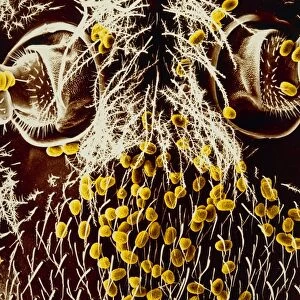
False-colour scanning electron micrograph (SEM) of pollen germinating on the stigma of the turnip flower, Brassica campestris. Of the many pollen grains which have lodged amongst the finger-like projections of the stigma, two have germinated & produced pollen tubes (centre right & bottom left). The pollen tube grows down inside the stigma, through the style (the column on which the stigma is supported) & enters the ovary at the base of the style; it protects & conveys two male gametes (sperm cells) to the female egg for fertilisation. Magnification: x400 at 6x4.5 cm, x250 at 35mm size. Red, pink & green
Science Photo Library features Science and Medical images including photos and illustrations
Media ID 6289271
© DR JEREMY BURGESS/SCIENCE PHOTO LIBRARY
Pollen Tube Re Production Reproductive Stigma Turnip Brassica Campestris
EDITORS COMMENTS
This print offers a mesmerizing glimpse into the intricate world of plant reproduction. Through the lens of a scanning electron microscope, we are granted access to the fascinating process of pollen germination on the stigma of a turnip flower. In this false-color image, we witness an array of pollen grains that have found their way onto the finger-like projections of the stigma. Amongst them, two exceptional grains have successfully germinated and produced delicate pollen tubes. These remarkable structures can be observed at the center-right and bottom-left sections of the photograph. As these pollen tubes grow downwards within the stigma, they traverse through a slender column known as the style. Eventually, they reach their destination: the ovary located at its base. Within these tubes lie two male gametes or sperm cells eagerly awaiting their opportunity to fertilize a female egg. The magnification power employed in capturing this breathtaking moment is truly impressive - x400 at 6x4.5 cm and x250 at 35mm size. The vibrant colors used in this image further enhance our appreciation for nature's beauty and complexity. Through this extraordinary visual representation provided by Science Photo Library, we gain insight into one small yet crucial aspect of plant life – pollination and reproduction – reminding us once again how intricately interconnected all living organisms truly are.
MADE IN THE UK
Safe Shipping with 30 Day Money Back Guarantee
FREE PERSONALISATION*
We are proud to offer a range of customisation features including Personalised Captions, Color Filters and Picture Zoom Tools
SECURE PAYMENTS
We happily accept a wide range of payment options so you can pay for the things you need in the way that is most convenient for you
* Options may vary by product and licensing agreement. Zoomed Pictures can be adjusted in the Basket.