Home > Popular Themes > Human Body
PET brain scans showing mistaken memory of words
![]()

Wall Art and Photo Gifts from Science Photo Library
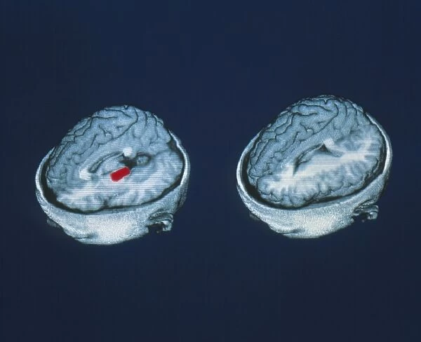
PET brain scans showing mistaken memory of words
Accurate memory of words. Coloured Positron Emis- sion Tomography (PET) scans of the brain showing accurate memory of words. Active areas of blood flow on the left-side of the brain are red. Women subjects listened to lists of words read aloud to them. Ten minutes later they were given lists to read containing the old plus a few new words. At left the hippocampus region is active remembering words. At right the temporoparietal region becomes active remembering those words that were spoken. This region distinguishes true memories from false memories (see photo P335/031) of previously spoken words. Active PET areas seen on a 3-D MRI scan
Science Photo Library features Science and Medical images including photos and illustrations
Media ID 6421879
© ERIC REIMAN, UNIVERSITY OF ARIZONA/SCIENCE PHOTO LIBRARY
Brain Function Brain Scan Memory Pet Scan Word Brain Mistaken Nervous System
EDITORS COMMENTS
This print showcases the intricate workings of the human brain when it comes to memory recall. The colored Positron Emission Tomography (PET) scans provide a visual representation of accurate memory of words in women subjects. The left-side of the brain, depicted in vibrant red, reveals active areas of blood flow within the hippocampus region. This area is responsible for accurately remembering words that were previously heard. On the right side, we observe activity in the temporoparietal region as it recalls those same spoken words. What makes this image truly fascinating is that it highlights how our brains distinguish between true memories and false ones. The temporoparietal region becomes engaged specifically when differentiating between what was actually said and what might have been mistakenly remembered. By studying these PET scans alongside a 3-D MRI scan, researchers gain valuable insights into brain function and memory processes. Understanding how our brains store and retrieve information can shed light on various aspects of human cognition. This remarkable photograph from Science Photo Library offers a glimpse into the complex world of neural connections and cognitive abilities. It serves as a reminder that even our most cherished memories may not always be as accurate as we believe them to be.
MADE IN THE UK
Safe Shipping with 30 Day Money Back Guarantee
FREE PERSONALISATION*
We are proud to offer a range of customisation features including Personalised Captions, Color Filters and Picture Zoom Tools
SECURE PAYMENTS
We happily accept a wide range of payment options so you can pay for the things you need in the way that is most convenient for you
* Options may vary by product and licensing agreement. Zoomed Pictures can be adjusted in the Basket.

