Home > Science > SEM
Oesophagus, SEM

![]()
Wall Art and Photo Gifts from Science Photo Library
Oesophagus, SEM
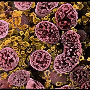
Oesophagus. Coloured scanning electron micrograph (SEM) of a freeze-fracture through the muscularis externa of the oesophagus. This forms part of the muscularis mucosa found throughout the gut. The upper part of the oesophagus contains striated muscle, which is muscle tissue characterised by strips. This muscle layer allows independent movements within the width of the mucous membrane. The oesophagus is a muscular tube about 25 centimetres long that runs from the back of the throat to the stomach. Magnification not known
Science Photo Library features Science and Medical images including photos and illustrations
Media ID 6449817
© STEVE GSCHMEISSNER/SCIENCE PHOTO LIBRARY
Alimentary Canal Digestion Digestive System Epithelial Fracture Gullet Keratinised Layered Layers Lining Mucosa Oesophageal Oesophagus Epithelium Squamous Stratified Striated Tissue Tract
FEATURES IN THESE COLLECTIONS
EDITORS COMMENTS
This print showcases the intricate details of the oesophagus, providing a glimpse into the fascinating world of human anatomy. The image, captured using a scanning electron microscope (SEM), reveals the layered and colourful structure of this vital organ. The muscularis externa, depicted in stunning detail, forms part of the muscularis mucosa that can be found throughout the entire digestive system. In particular, the upper portion of the oesophagus features striated muscle tissue characterized by its distinctive striped appearance. This unique muscle layer enables independent movements within the width of the mucous membrane. Stretching approximately 25 centimetres from throat to stomach, this muscular tube plays a crucial role in our digestion process. Its lining consists of squamous epithelial cells arranged in stratified and cornified layers. Notably, some areas exhibit keratinization while others remain non-keratinized. As we marvel at this remarkable SEM image, we are reminded once again of our body's extraordinary complexity and resilience. It serves as a reminder that even on a microscopic level, every component works harmoniously to ensure our overall well-being. This awe-inspiring photograph is brought to you by Science Photo Library – an esteemed source for scientific imagery that continues to inspire curiosity and understanding about our incredible human form.
MADE IN THE UK
Safe Shipping with 30 Day Money Back Guarantee
FREE PERSONALISATION*
We are proud to offer a range of customisation features including Personalised Captions, Color Filters and Picture Zoom Tools
SECURE PAYMENTS
We happily accept a wide range of payment options so you can pay for the things you need in the way that is most convenient for you
* Options may vary by product and licensing agreement. Zoomed Pictures can be adjusted in the Basket.