Home > Popular Themes > Human Body
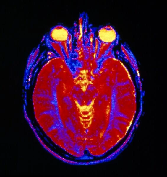
NMR scan of head, axial section taken at eye level
![]()

Wall Art and Photo Gifts from Science Photo Library
NMR scan of head, axial section taken at eye level
False-colour Magnetic Resonance Image (MRI) of an axial section through a human head, showing the division of the brain into left & right cerebral hemispheres. MRI (also called NMR imaging) distinguishes between the grey & white matter components of the brain. Here, outer grey matter is coloured red, and the inner white matter coloured blue. This scan was made at eye-level - vitreous humour inside each eye is coloured yellow. The layer of fat under the skin of the scalp appears red & purple. Bone of the skull and the cerebro-spinal fluid which bathes the brain appear black
Science Photo Library features Science and Medical images including photos and illustrations
Media ID 6421620
© CNRI/SCIENCE PHOTO LIBRARY
EDITORS COMMENTS
This print showcases a false-colour Magnetic Resonance Image (MRI) of an axial section through a human head, providing a remarkable glimpse into the intricate division of the brain. The left and right cerebral hemispheres are beautifully highlighted, allowing us to appreciate the complexity and interconnectedness of our most vital organ. Through the power of MRI technology, this image distinguishes between the grey matter, responsible for processing information in our brains, and the white matter that facilitates communication between different regions. The outer grey matter is vividly displayed in striking red hues, while the inner white matter is elegantly depicted in shades of mesmerizing blue. At eye level within this scan lies another fascinating detail - the vitreous humour inside each eye takes on a vibrant yellow hue. This serves as a reminder that even within such advanced imaging techniques, we can still observe elements unique to our anatomy. Furthermore, this image offers additional insights into other components surrounding our brain. The layer of fat beneath the scalp appears as an intriguing blend of red and purple tones. Meanwhile, bone structures comprising the skull stand out prominently against a contrasting black background alongside cerebro-spinal fluid which bathes and protects our precious brain. In essence, this NMR scan captures both scientific precision and artistic beauty simultaneously. It invites us to marvel at one of nature's greatest wonders -the human nervous system- while showcasing how modern technology enables us to explore its intricacies with unparalleled clarity.
MADE IN THE UK
Safe Shipping with 30 Day Money Back Guarantee
FREE PERSONALISATION*
We are proud to offer a range of customisation features including Personalised Captions, Color Filters and Picture Zoom Tools
SECURE PAYMENTS
We happily accept a wide range of payment options so you can pay for the things you need in the way that is most convenient for you
* Options may vary by product and licensing agreement. Zoomed Pictures can be adjusted in the Basket.

