Home > Popular Themes > Human Body
Mouth anatomy, artwork

![]()
Wall Art and Photo Gifts from Science Photo Library
Mouth anatomy, artwork
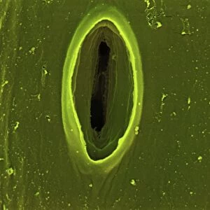
Mouth anatomy. Artwork of a section through the human head showing the anatomy of the mouth, nose and upper throat. Above the mouth is the nasal cavity (black), an air passage that connects with the top of the throat (pharynx). The nasal cavity is separated from the oral cavity by the hard (front) and soft (back) palate. The oral cavity is dominated by the tongue (red, bottom centre). Inhaled air is filtered, warmed and moistened by the mucous membrane of the nasal cavity and passes through the pharynx to the trachea (windpipe, bottom left). Behind the trachea is the oesophagus, which carries food to the stomach to be digested
Science Photo Library features Science and Medical images including photos and illustrations
Media ID 6344487
© FRANCIS LEROY, BIOCOSMOS/SCIENCE PHOTO LIBRARY
Mandible Mouth Nasal Cavity Nose Oral Cavity Pharynx Profile Saggital Teeth Throat Tongue Section
EDITORS COMMENTS
This artwork captures the intricate details of the mouth anatomy, providing a fascinating glimpse into the inner workings of the human head. Against a striking black background, this print showcases a section through the head, revealing not only the mouth but also the nose and upper throat. The nasal cavity takes center stage in this composition, depicted in bold black lines. It serves as an air passage that connects with the top of the throat known as pharynx. The hard palate at the front and soft palate at the back separate it from oral cavity dominated by a vibrant red tongue positioned at its bottom center. Inhaled air undergoes filtration, warming, and moistening within this mucous membrane-lined nasal cavity before passing through to reach trachea or windpipe situated towards bottom left. Behind it lies another crucial structure - esophagus - responsible for transporting food to be digested in our stomach. With meticulous attention to detail, this illustration beautifully highlights various elements such as teeth, jawbone (mandible), and even provides a side view cut-out for enhanced understanding. This anatomical masterpiece is both educational and visually stunning; an ideal addition for anyone interested in biology or simply fascinated by how our bodies function.
MADE IN THE UK
Safe Shipping with 30 Day Money Back Guarantee
FREE PERSONALISATION*
We are proud to offer a range of customisation features including Personalised Captions, Color Filters and Picture Zoom Tools
SECURE PAYMENTS
We happily accept a wide range of payment options so you can pay for the things you need in the way that is most convenient for you
* Options may vary by product and licensing agreement. Zoomed Pictures can be adjusted in the Basket.