Home > Popular Themes > Human Body
Large intestine
![]()

Wall Art and Photo Gifts from Science Photo Library
Large intestine
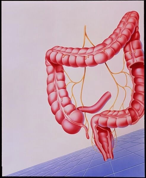
Colon. Artwork showing the human colon (large intestine). The yellow fibres represent nerves, which control the involuntary muscular movements (peristalsis) of the colon. The colon has no digestive function but absorbs water and electro- lytes from the residue of digestion. It consists of 4 sections: the ascending (left), transverse (top), descending (right, cut away) and sigmoid (bottom). The sigmoid colon joins with the rectum. Three bands of longitudinal muscle, the teniae coli, run the length of the organ, from the appendix (tail-like at bottom left) to the rectum. The teniae help to form haustra, the compartment- like pouches along the length of the colon
Science Photo Library features Science and Medical images including photos and illustrations
Media ID 6422782
© JOHN BAVOSI/SCIENCE PHOTO LIBRARY
Appendix Artwor Colon Digestive System Innervation Intestine Large Large Intestine Nerve Nerves
EDITORS COMMENTS
This artwork showcases the intricate beauty of the human colon, also known as the large intestine. The vibrant yellow fibers depicted in this print represent the nerves that play a crucial role in controlling the involuntary muscular movements of the colon, known as peristalsis. While it does not participate in digestion, this vital organ absorbs water and electrolytes from digested residue. The large intestine is divided into four sections: ascending (left), transverse (top), descending (right, cut away), and sigmoid (bottom). The sigmoid colon seamlessly connects with the rectum to facilitate waste elimination. Along its length, three bands of longitudinal muscle called teniae coli can be observed. These muscles extend from the appendix at bottom left all the way to the rectum and aid in forming haustra – compartment-like pouches that contribute to efficient functioning. This mesmerizing illustration provides a comprehensive view of both anatomical structure and physiological processes within our digestive system. It highlights how innervation plays a pivotal role in ensuring proper functioning of our large intestine. This remarkable artwork serves as a testament to Science Photo Library's commitment to providing educational resources that showcase scientific wonders while capturing their artistic essence.
MADE IN THE UK
Safe Shipping with 30 Day Money Back Guarantee
FREE PERSONALISATION*
We are proud to offer a range of customisation features including Personalised Captions, Color Filters and Picture Zoom Tools
SECURE PAYMENTS
We happily accept a wide range of payment options so you can pay for the things you need in the way that is most convenient for you
* Options may vary by product and licensing agreement. Zoomed Pictures can be adjusted in the Basket.

