Home > Popular Themes > Human Body
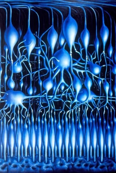
Illustration of nerve cells in mammalian retina
![]()

Wall Art and Photo Gifts from Science Photo Library
Illustration of nerve cells in mammalian retina
Illustration of the layers of nerve cells comprising the mammalian retina, the light- sensitive layer of the eye. At bottom, the outermost layer of pigmented cells supports a number of rod- & cone-type photosensitive cells. The rods outnumber cones by about 20:1 - hence only cones are shown here. The cell bodies above the rods & cones layer form the integrating bipolar cell layer; a single cell body may be seen to serve several rod receptors via numerous synapses (junctions). The upper ganglion layer of cell bodies belong to fibres comprising the optic nerve (top)
Science Photo Library features Science and Medical images including photos and illustrations
Media ID 6422316
© FRANCIS LEROY, BIOCOSMOS/SCIENCE PHOTO LIBRARY
EDITORS COMMENTS
This print showcases the intricate and awe-inspiring world of nerve cells in the mammalian retina. The layers of these nerve cells, which make up the light-sensitive layer of our eyes, are beautifully illustrated here. Starting at the bottom, we can see a layer of pigmented cells that provide support to numerous rod and cone-type photosensitive cells. Interestingly, cones are exclusively depicted in this illustration as they outnumber rods by about 20 to 1. These cones play a crucial role in our ability to perceive color and detailed vision. Moving upward, we encounter the integrating bipolar cell layer where cell bodies connect with multiple rod receptors through countless synapses or junctions. This intricate network allows for efficient transmission of visual information from the photoreceptor cells. Finally, at the topmost layer, we observe cell bodies belonging to fibers that form the optic nerve. These ganglion cells serve as messengers carrying visual signals from our eyes to our brain for interpretation. The complexity and precision displayed within this artwork remind us of how remarkable our visual sense truly is. It serves as a testament to both scientific understanding and artistic representation merging seamlessly together. This stunning image from Science Photo Library not only highlights the beauty found within our own anatomy but also sparks curiosity about how we perceive sight on a daily basis.
MADE IN THE UK
Safe Shipping with 30 Day Money Back Guarantee
FREE PERSONALISATION*
We are proud to offer a range of customisation features including Personalised Captions, Color Filters and Picture Zoom Tools
SECURE PAYMENTS
We happily accept a wide range of payment options so you can pay for the things you need in the way that is most convenient for you
* Options may vary by product and licensing agreement. Zoomed Pictures can be adjusted in the Basket.

