Home > Science > SEM
False-colour SEM of sperm on uterine surface

![]()
Wall Art and Photo Gifts from Science Photo Library
False-colour SEM of sperm on uterine surface
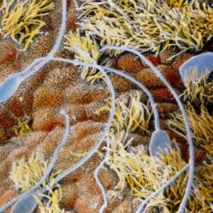
Capacitation of spermatozoa. False-colour scanning electron micrograph of a group of spermatozoa (turquoise) floating over the endometrium, the internal wall of the uterus. Thanks to their movements through the ciliated epithelium (yellow) spermatozoa undergo the process known as capacitation. During this phase, which lasts about seven hours, a glycoprotein coat and seminal plasma proteins are removed from the heads of spermatozoa. This enables a spermatozoon, when close to the egg, to release special enzymes which allows it to penetrate the surrounding membranes of the egg. Magnification: x2460 at 6x7cm size
Science Photo Library features Science and Medical images including photos and illustrations
Media ID 6454805
© PROFESSORS P.M. MOTTA & J. VAN BLERKOM/ SCIENCE PHOTO LIBRARY
Endometrium Re Production Reproductive System Sperm Spermatozoa Uterus False Coloured
FEATURES IN THESE COLLECTIONS
EDITORS COMMENTS
This print showcases the intricate process of capacitation in spermatozoa. In this false-colour scanning electron micrograph, a group of spermatozoa can be seen floating over the endometrium, the internal wall of the uterus. The turquoise hue beautifully highlights these tiny cells as they undergo a crucial transformation. Capacitation is a vital phase lasting approximately seven hours, during which glycoprotein coats and seminal plasma proteins are stripped away from the heads of spermatozoa. This preparatory step enables a spermatozoon to release special enzymes when near an egg, allowing it to penetrate the surrounding membranes with precision and accuracy. The yellow ciliated epithelium visible in this image plays a significant role in facilitating this process by aiding the movement of sperm through its microscopic channels. Through their synchronized movements within this complex environment, these remarkable cells embark on their journey towards fertilization. With magnification at x2460 and printed at 6x7cm size, every detail becomes apparent - highlighting both the intricacy and beauty found within our reproductive system. This stunning photograph not only provides valuable insights into human reproduction but also serves as a testament to the wonders that occur within our bodies on such minuscule scales.
MADE IN THE UK
Safe Shipping with 30 Day Money Back Guarantee
FREE PERSONALISATION*
We are proud to offer a range of customisation features including Personalised Captions, Color Filters and Picture Zoom Tools
SECURE PAYMENTS
We happily accept a wide range of payment options so you can pay for the things you need in the way that is most convenient for you
* Options may vary by product and licensing agreement. Zoomed Pictures can be adjusted in the Basket.