Home > Popular Themes > Human Body
Facial muscles and internal structure of the head

![]()
Wall Art and Photo Gifts from Science Photo Library
Facial muscles and internal structure of the head
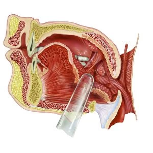
Two models showing the arrangement of the facial and neck muscles (left) and a midsagittal section of the head and neck (right). The sectioned model shows the cerebrum, the largest part of the brain, in yellow and the brainstem, the lower part of the brain, which connects the brain to the spinal cord. The largest tube at bottom right is the trachea and the narrower tube behind it the oesophagus. The model on the left shows the location of the muscle bundles around the eyes and mouth and also the muscles in the neck that control the movements of the head. On this model the parotid gland is found to the right of the ear
Science Photo Library features Science and Medical images including photos and illustrations
Media ID 6452705
© GEOFF TOMPKINSON/SCIENCE PHOTO LIBRARY
Face Neck Oral Cavity Sagittal Section Brain
EDITORS COMMENTS
This print showcases the intricate details of the facial muscles and internal structure of the head. Two models are featured, each shedding light on different aspects of human anatomy. On the left, we observe a model that highlights the arrangement of facial and neck muscles. The muscle bundles surrounding the eyes and mouth are clearly visible, emphasizing their crucial role in our expressions and movements. Additionally, this model reveals the neck muscles responsible for controlling head movements. On the right side, a midsagittal section of the head and neck is displayed. This sectioned model provides an insightful view into our brain's composition. The cerebrum, depicted in vibrant yellow hues, represents its largest part while below it lies the brainstem - connecting our brain to our spinal cord. The complexity of human physiology becomes even more apparent as we shift our attention to other structures within this image. Located at bottom right is a large tube known as trachea or windpipe which allows air passage into our lungs; behind it lies a narrower tube called oesophagus through which food travels to reach our stomach. Intriguingly, this print also highlights an important gland - parotid gland - positioned to the right of one's ear. Overall, this remarkable visual representation serves as a testament to both scientific curiosity and appreciation for human body intricacies captured by Science Photo Library without any commercial intentions involved
MADE IN THE UK
Safe Shipping with 30 Day Money Back Guarantee
FREE PERSONALISATION*
We are proud to offer a range of customisation features including Personalised Captions, Color Filters and Picture Zoom Tools
SECURE PAYMENTS
We happily accept a wide range of payment options so you can pay for the things you need in the way that is most convenient for you
* Options may vary by product and licensing agreement. Zoomed Pictures can be adjusted in the Basket.