Home > Popular Themes > Human Body
Eye structure, light micrograph

![]()
Wall Art and Photo Gifts from Science Photo Library
Eye structure, light micrograph
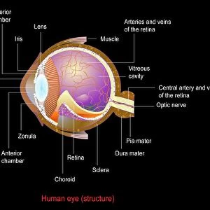
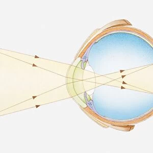
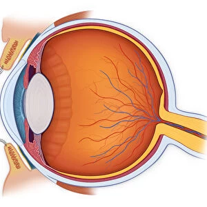
Eye structure. Coloured light micrograph of a section through the inner surface of a human eye. This slice shows the vitreous humour (transparent liquid in the eye, top), the retina (pink), the choroid (black layer) and the sclera (white external surface, bottom). The eye works by allowing light to be focused by the lens onto the retina. The retina contains photoreceptor cells (small purple dots), which allow the eye to distinguish between colours (cone cells) and to see at night (rod cells). The choroid layer lines the inside of the eye underneath the retina and is pigmented to prevent light reflecting inside the eye and distorting the image. Magnification: x160 when printed at 10cm wide
Science Photo Library features Science and Medical images including photos and illustrations
Media ID 6422358
© STEVE GSCHMEISSNER/SCIENCE PHOTO LIBRARY
Choroid Cross Section Eye Ball False Colour Histological Histology Layers Longitudinal Nerve Fibres Photoreceptors Retina Retinal Sclera Sense Sight Slice Tissue Tissues Vision Visual Cells False Coloured Light Micrograph Light Microscope Section
EDITORS COMMENTS
This print showcases the intricate structure of the human eye, revealing its remarkable ability to perceive and interpret light. In this coloured light micrograph, we are granted a glimpse into the inner surface of an eye, exposing its various layers and components. At first glance, we observe the vitreous humour, a transparent liquid that fills the top portion of the eye. Below it lies the retina, painted in a delicate shade of pink. This vital layer contains photoreceptor cells represented by small purple dots; these cells enable us to distinguish between colours (cone cells) and see in low-light conditions (rod cells). The black layer known as choroid lines the inside of the eye beneath the retina. Its pigmentation serves as a shield against internal reflections that could distort our vision. Finally, at the bottom rests the sclera – an external white surface that encompasses and protects this extraordinary organ. Through meticulous magnification at 160 times its actual size when printed at 10cm wide, Science Photo Library has captured not only anatomical precision but also conveyed a sense of wonder within this image. It reminds us just how intricately woven our visual system is and highlights both its biological complexity and aesthetic beauty. This photograph offers valuable insights into sight biology while celebrating one's appreciation for their own healthy vision – an awe-inspiring reminder of nature's brilliance within each individual's eyesight.
MADE IN THE UK
Safe Shipping with 30 Day Money Back Guarantee
FREE PERSONALISATION*
We are proud to offer a range of customisation features including Personalised Captions, Color Filters and Picture Zoom Tools
SECURE PAYMENTS
We happily accept a wide range of payment options so you can pay for the things you need in the way that is most convenient for you
* Options may vary by product and licensing agreement. Zoomed Pictures can be adjusted in the Basket.