Diarrhoea, artwork

![]()
Wall Art and Photo Gifts from Science Photo Library
Diarrhoea, artwork
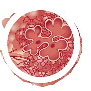
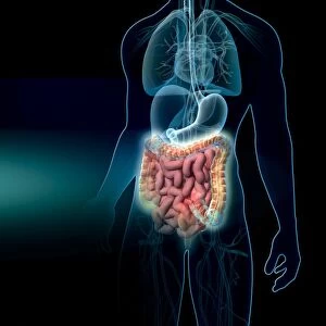
Diarrhoea. Artwork of the human large intestine surrounded by some of the microorganisms that can cause diarrhoea. Diarrhoea is the frequent passing of liquid stools as a symptom of inflammation, infection or even anxiety. Ingested microorganisms as a result of poor hygiene or food storage can lead to various diseases whose symptoms include diarrhoea. Rod-shaped (bacillus) bacteria are seen at upper left, upper and center right, which could include Salmonella, Escherichia coli (E. coli), Campylobacter and Cryptosporidium. The protozoan Giardia lamblia is seen at bottom right, Staphylococcus bacteria at centre left and Vibrio cholerae bacteria at lower left
Science Photo Library features Science and Medical images including photos and illustrations
Media ID 6422263
© JOHN BAVOSI/SCIENCE PHOTO LIBRARY
Anus Bacillus Bacteria Bacterium Bowel Colon Diarrhoea Digestion Escherichia Coli Intestinal Large Intestine Protozoa Rectum Salmonella Symptom Symptoms Vertical Vibrio Cholerae Condition Disorder Health Care
EDITORS COMMENTS
This print titled "Diarrhoea, artwork" showcases the intricate details of the human large intestine surrounded by a multitude of microorganisms that can cause diarrhoea. Diarrhoea is a distressing condition characterized by frequent passing of liquid stools, often stemming from inflammation, infection, or even anxiety. This thought-provoking artwork sheds light on how ingesting microorganisms due to poor hygiene or food storage practices can lead to various diseases with diarrhoea as a prominent symptom. The image highlights different types of bacteria and protozoa responsible for causing diarrhoeal illnesses. Rod-shaped (bacillus) bacteria occupy the upper left, upper and center right sections, potentially including notorious culprits like Salmonella, Escherichia coli (E. coli), Campylobacter, and Cryptosporidium. The bottom right corner reveals the presence of Giardia lamblia protozoan while Staphylococcus bacteria are depicted at center left and Vibrio cholerae bacteria at lower left. Through this artistic representation blending science and creativity seamlessly together, viewers gain insight into the complex world within our digestive system where these microorganisms wreak havoc when conditions are unfavorable. It serves as a reminder of the importance of maintaining proper hygiene practices and safe food storage methods to prevent such debilitating symptoms associated with bowel disorders. This remarkable print from Science Photo Library not only educates but also sparks curiosity about medical illustrations in understanding health-related issues concerning our intestines' well-being – an essential aspect of overall healthcare management.
MADE IN THE UK
Safe Shipping with 30 Day Money Back Guarantee
FREE PERSONALISATION*
We are proud to offer a range of customisation features including Personalised Captions, Color Filters and Picture Zoom Tools
SECURE PAYMENTS
We happily accept a wide range of payment options so you can pay for the things you need in the way that is most convenient for you
* Options may vary by product and licensing agreement. Zoomed Pictures can be adjusted in the Basket.