Coloured X-ray of fractured shin bone (tibia)
")
![]()
Wall Art and Photo Gifts from Science Photo Library
Coloured X-ray of fractured shin bone (tibia)
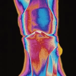
Broken leg. Coloured X-ray showing a fracture of the shin bone (tibia) of the leg. The tibia is the larger of the two bones of the lower leg, with the injury seen at centre right. Superimposed are drawn yellow lines marked 1 & 2 measuring length of each tibia bone. The fractured tibia marked 1 has lost considerable length. Treatment of fractures involves moving the broken ends into alignment, and immobilising the leg while the bones reunite. Because the tibia is a weight- bearing bone it can take up to six months to fully heal. Shortening of one limb may require a walking aid such as specialized shoes. An X-ray to measure bone length is called a topogram
Science Photo Library features Science and Medical images including photos and illustrations
Media ID 6424249
© MEHAU KULYK/SCIENCE PHOTO LIBRARY
Broken Broken Leg Fracture Injury Tibia Condition Disorder Health Care
EDITORS COMMENTS
This print from Science Photo Library showcases a coloured X-ray of a fractured shin bone (tibia), providing an insightful glimpse into the world of medical imaging. The image vividly displays a broken leg, with the fracture clearly visible at the center right. To aid in understanding the severity of the injury, yellow lines marked 1 and 2 have been superimposed to measure the length of each tibia bone. The significance of this particular X-ray lies in its ability to highlight both the condition and treatment process for fractures. When faced with such injuries, medical professionals must carefully realign and immobilize the broken ends to facilitate healing. However, due to its weight-bearing nature, complete recovery for a fractured tibia can take up to six months. Notably, this X-ray reveals that one limb has experienced considerable shortening as a result of the fracture. In cases like these, specialized shoes or walking aids may be necessary to restore balance and mobility. Furthermore, it is worth mentioning that an X-ray used specifically for measuring bone length is referred to as a topogram—a valuable tool in assessing damage and planning appropriate treatment strategies. Overall, this visually striking print serves as a testament not only to advancements in medical technology but also highlights how healthcare professionals strive tirelessly towards restoring health and well-being even amidst challenging circumstances.
MADE IN THE UK
Safe Shipping with 30 Day Money Back Guarantee
FREE PERSONALISATION*
We are proud to offer a range of customisation features including Personalised Captions, Color Filters and Picture Zoom Tools
SECURE PAYMENTS
We happily accept a wide range of payment options so you can pay for the things you need in the way that is most convenient for you
* Options may vary by product and licensing agreement. Zoomed Pictures can be adjusted in the Basket.