Home > Science > SEM
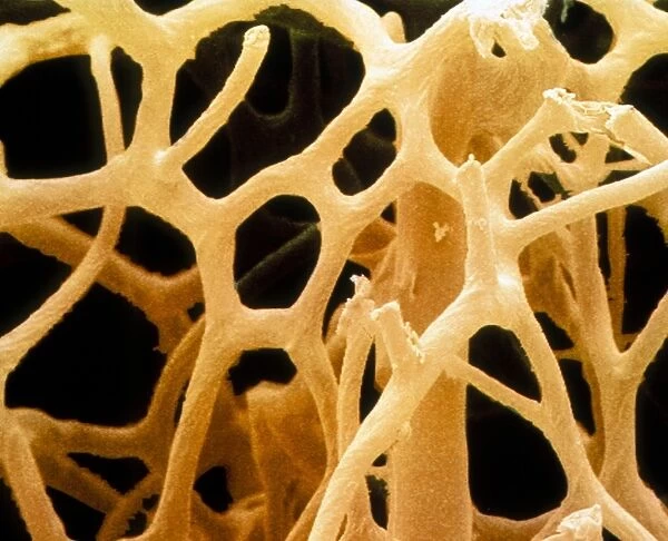
Coloured SEM of a natural sponge
![]()

Wall Art and Photo Gifts from Science Photo Library
Coloured SEM of a natural sponge
Sponge. Coloured scanning electron micrograph (SEM) of an unidentified sponge, phylum Porifera. The branching structure of the sponges body is supported by an internal skeleton of calcareous or siliceous spicules (spines) and fibres of the protein spongin. Sponges are sessile multicellular animals which are primitively differentiated; the body wall consisting of just two layers. Water canals link pores on the surface with a communal exhalent tube. All sponges are current or filter feeders. The water current created by flagellated cells carries particles and tiny organisms into the sponges body. Magnification: x120 at 6x7cm size. x296 at 8x6, x158 at 10x7cm master size
Science Photo Library features Science and Medical images including photos and illustrations
Media ID 9306957
© POWER AND SYRED/SCIENCE PHOTO LIBRARY
Porifera Poriferan Spicule Spicules Sponge
EDITORS COMMENTS
This print showcases the intricate beauty of a natural sponge, captured through a coloured scanning electron microscope (SEM). Belonging to the phylum Porifera, this unidentified sponge exhibits a remarkable branching structure that is supported by an internal skeleton composed of calcareous or siliceous spicules and protein spongin fibers. Sponges are fascinating sessile multicellular animals characterized by their primitive differentiation, with just two layers comprising their body wall. Water canals intricately connect pores on the surface of the sponge to a communal exhalent tube. This unique feature allows all sponges to function as current or filter feeders. The water current, generated by flagellated cells within the sponge's body, carries particles and tiny organisms into its interior. At 6x7cm size magnification, this SEM image offers us a closer look at this mesmerizing creature at x120 magnification. For larger sizes such as 8x6cm and 10x7cm master size prints, higher levels of detail emerge at x296 and x158 magnifications respectively. Nature enthusiasts and zoology lovers will undoubtedly appreciate this extraordinary glimpse into the world of invertebrates. Taken during the Challenger Expedition in 1874, this photograph from Science Photo Library serves as both an educational tool for studying poriferans and an awe-inspiring testament to wildlife's diversity.
MADE IN THE UK
Safe Shipping with 30 Day Money Back Guarantee
FREE PERSONALISATION*
We are proud to offer a range of customisation features including Personalised Captions, Color Filters and Picture Zoom Tools
SECURE PAYMENTS
We happily accept a wide range of payment options so you can pay for the things you need in the way that is most convenient for you
* Options may vary by product and licensing agreement. Zoomed Pictures can be adjusted in the Basket.


