Home > Popular Themes > Human Body
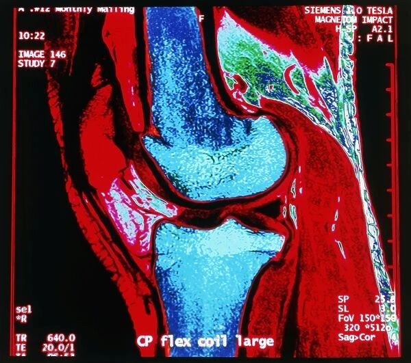
Coloured MRI scan of human knee joint, side view
![]()

Wall Art and Photo Gifts from Science Photo Library
Coloured MRI scan of human knee joint, side view
Knee joint. Coloured Magnetic Resonance Imaging (MRI) scan of a sagittal section through a human knee joint. Two bones (coloured blue) meet at the knee, forming a hinge-joint. At top is the femur (thigh-bone), which articulates with the tibia (shin-bone) at bottom. The patella or kneecap (red, located at upper left) is a protective bone at the front of the knee held in position by muscles and tendons. Two discs of protective cartilage cover the surfaces of the femur and tibia to reduce friction between these bones. The round bottom surface of the femur forms a narrow ridge that moves through a groove in the tibia. This allows hinge movement, with slight rotation
Science Photo Library features Science and Medical images including photos and illustrations
Media ID 6419942
© GEOFF TOMPKINSON/SCIENCE PHOTO LIBRARY
Bones Femur Joint Knee Knee Cap Knee Joint Mri Scan Patella Tibia Hinge Joint
EDITORS COMMENTS
This print showcases a coloured MRI scan of the human knee joint, providing a detailed side view. The image highlights the intricate structure and functionality of this vital joint. Two bones, depicted in striking blue hues, elegantly meet at the knee to form a hinge-joint. At the top of the frame lies the femur or thigh-bone, which articulates with the tibia or shin-bone positioned at the bottom. A prominent feature captured in vibrant red is the patella or kneecap, situated towards the upper left corner. This protective bone safeguards the front of our knees and remains securely held in place by muscles and tendons. To ensure smooth movement between these bones, two discs composed of protective cartilage cover their surfaces. These discs play an essential role in reducing friction during motion within this weight-bearing joint. Notably, we can observe that as part of its design for flexibility and stability, there is a narrow ridge on the round bottom surface of femur that glides through a groove present in tibia. This mechanism enables hinge-like movements while allowing for slight rotation as well. Overall, this mesmerizing snapshot provides valuable insights into both normal anatomy and skeletal dynamics associated with our knee joints – an integral component contributing to our ability to walk, run and perform various physical activities effortlessly.
MADE IN THE UK
Safe Shipping with 30 Day Money Back Guarantee
FREE PERSONALISATION*
We are proud to offer a range of customisation features including Personalised Captions, Color Filters and Picture Zoom Tools
SECURE PAYMENTS
We happily accept a wide range of payment options so you can pay for the things you need in the way that is most convenient for you
* Options may vary by product and licensing agreement. Zoomed Pictures can be adjusted in the Basket.

