Home > Popular Themes > Human Body
Bones of the leg

![]()
Wall Art and Photo Gifts from Science Photo Library
Bones of the leg
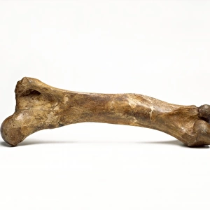
Bones of the leg. Artwork of human femurs (thigh bones) and patellae (kneecaps), taken from anatomist William Cheseldens textbook Osteographia, or the Anatomy of the Bones, published in 1733. The right femur is shown at the front and the left behind. The balls at the top of the femurs fit into the hip sockets. At the bottom are the upper halves of the hinge joints that form the knees. The patellae protect the front of the knee joints
Science Photo Library features Science and Medical images including photos and illustrations
Media ID 6419496
© MEHAU KULYK/SCIENCE PHOTO LIBRARY
1700s 18th Century Drawing Femoral Femur Femurs Historical Image Imagery Knee Knee Cap Patella Skeletal Thigh 1733 Mono Chrome
EDITORS COMMENTS
This print showcases the intricate beauty of human anatomy through a historical lens. Taken from William Cheselden's renowned textbook "Osteographia, or the Anatomy of the Bones" published in 1733, it offers a glimpse into the detailed study of bones during that era. The image features two human femurs (thigh bones) and patellae (kneecaps), elegantly captured in monochrome. The right femur takes center stage at the front, while its counterpart stands gracefully behind. These essential leg bones are responsible for supporting our body weight and facilitating movement. At the top of each femur, we observe spherical structures that fit snugly into hip sockets, enabling fluid motion within our pelvis. Towards the bottom, we encounter upper halves of hinge joints forming our knees – crucial connectors between thigh and lower leg bones. Notably present are patellae, protecting the front part of these knee joints. Their presence ensures stability and safeguards against potential injuries during physical activities. This artwork not only serves as an invaluable resource for biology enthusiasts but also provides a fascinating window into medical history. It represents an era when anatomical knowledge was meticulously documented to advance scientific understanding. With its blend of artistry and scientific precision, this historical illustration invites us to marvel at both the complexity and elegance inherent in every aspect of our skeletal structure - an enduring testament to nature's remarkable design.
MADE IN THE UK
Safe Shipping with 30 Day Money Back Guarantee
FREE PERSONALISATION*
We are proud to offer a range of customisation features including Personalised Captions, Color Filters and Picture Zoom Tools
SECURE PAYMENTS
We happily accept a wide range of payment options so you can pay for the things you need in the way that is most convenient for you
* Options may vary by product and licensing agreement. Zoomed Pictures can be adjusted in the Basket.