Home > Popular Themes > Human Body
Atrial fibrillation, artwork
![]()

Wall Art and Photo Gifts from Science Photo Library
Atrial fibrillation, artwork
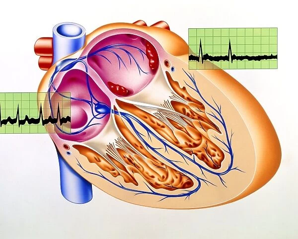
Atrial fibrillation. Artwork of a section through a human heart during atrial fibrillation, a rapid, irregular heartbeat. Two electrocardiogram traces (ECGs) are seen at centre left and upper right. The ECGs show the irregularity of the heart rhythm (arrhythmia). The electrical impulses that coordinate heart contractions start at the sinoatrial node (blue, upper left) and run down to the atrioventricular node (blue, centre left). When the atria (upper heart chambers, pink) contract irregularly & too rapidly, the ventricles (lower heart chambers, orange) have trouble keeping pace. ECGs are used to diagnose heart conditions such as arrhythmia
Science Photo Library features Science and Medical images including photos and illustrations
Media ID 6422671
© JOHN BAVOSI/SCIENCE PHOTO LIBRARY
Arrhythmia Cardiac Electrocardiography Heart Beat Horizontal Irregular Irregularity Rhythm Sinoatrial Node Condition Disorder Health Care
EDITORS COMMENTS
This artwork captures the complexity of atrial fibrillation, a condition characterized by a rapid and irregular heartbeat. The image provides an intricate view of a section through a human heart during this arrhythmia, showcasing the inner workings of our most vital organ. At the center left and upper right, two electrocardiogram traces (ECGs) vividly display the erratic nature of the heart rhythm affected by atrial fibrillation. These ECGs serve as diagnostic tools in identifying various heart conditions, including arrhythmias. Originating from the sinoatrial node depicted in blue at the upper left corner, electrical impulses travel down to reach the atrioventricular node situated at the center left. The pink-colored chambers represent the atria or upper heart chambers responsible for contracting irregularly and too rapidly during this disorder. Consequently, this puts strain on their counterparts below –the ventricles shown in orange– which struggle to maintain synchronization with such chaotic contractions. This thought-provoking illustration not only showcases medical artistry but also serves as an educational tool for healthcare professionals and patients alike. By shedding light on atrial fibrillation's intricacies within our own bodies, it emphasizes both its impact on cardiac health and highlights how electrocardiography plays a crucial role in diagnosis. Science Photo Library has once again captured science's beauty through visual representation while providing valuable insights into cardiovascular health.
MADE IN THE UK
Safe Shipping with 30 Day Money Back Guarantee
FREE PERSONALISATION*
We are proud to offer a range of customisation features including Personalised Captions, Color Filters and Picture Zoom Tools
SECURE PAYMENTS
We happily accept a wide range of payment options so you can pay for the things you need in the way that is most convenient for you
* Options may vary by product and licensing agreement. Zoomed Pictures can be adjusted in the Basket.

