Home > Popular Themes > Human Body
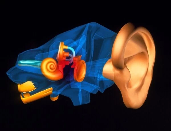
3-D computer model of the anatomy of the human ear
![]()

Wall Art and Photo Gifts from Science Photo Library
3-D computer model of the anatomy of the human ear
Ear anatomy. Three-dimensional computer model of the anatomy of the human ear. Bone is dark blue. At right, the outer ear pinna collects sound waves which pass down the auditory canal to strike the flat eardrum (at centre). Vibrations of sound are transmitted across three tiny bones in the middle ear which touch the eardrum (not all these bones are seen). Sound passes to structures of the inner ear (centre left). These are the semi-circular canals with three coils which detect balance. And the spiral-shaped cochlea containing hair cells that register sound as nerve impulses. Impulses activate the auditory nerve (light blue) which transmits the sound to the brain
Science Photo Library features Science and Medical images including photos and illustrations
Media ID 6422404
© PASIEKA/SCIENCE PHOTO LIBRARY
Artwk Auditory Sense Cochlea Computer Graphic Hearing Inner Ear Middle Ear Semicircular Canal
EDITORS COMMENTS
This print showcases a remarkable 3-D computer model of the intricate anatomy of the human ear. The image beautifully depicts the various components that make up this vital sensory organ. The bones, depicted in a dark blue hue, form the foundation of the ear's structure. At the right side of the image, we see the outer ear pinna, which acts as a collector for sound waves. These sound waves then travel down through the auditory canal until they reach and strike against the flat eardrum located at its center. This interaction creates vibrations that are transmitted across three tiny bones within the middle ear (although not all these bones are visible in this particular representation). Moving towards structures within the inner ear on our left-hand side, we encounter an intriguing sight - semi-circular canals with their distinctive three coils responsible for detecting balance. Adjacent to them lies another crucial component: a spiral-shaped cochlea housing hair cells that register sound as nerve impulses. The culmination of this process occurs when these impulses activate and stimulate our auditory nerve represented by light blue lines in this artwork. Finally, it is through this pathway that sounds are transmitted to our brain for interpretation and perception. This visually stunning piece from Science Photo Library provides us with an awe-inspiring glimpse into one of nature's most fascinating creations –the human ear– showcasing both its complexity and beauty simultaneously.
MADE IN THE UK
Safe Shipping with 30 Day Money Back Guarantee
FREE PERSONALISATION*
We are proud to offer a range of customisation features including Personalised Captions, Color Filters and Picture Zoom Tools
SECURE PAYMENTS
We happily accept a wide range of payment options so you can pay for the things you need in the way that is most convenient for you
* Options may vary by product and licensing agreement. Zoomed Pictures can be adjusted in the Basket.

