Fraud Blocker Skip to main content

Home > Popular Themes > DNA

DNA Collection (page 23)

3,051 items

Background imageDNA Collection: Notch transcription, molecular model

Notch transcription, molecular model
Notch transcription. Molecular model showing a strand of DNA (deoxyribonucleic acid) being acted upon by various proteins and other molecules

Background imageDNA Collection: Mitochondrial structure, artwork C015 / 6765

Mitochondrial structure, artwork C015 / 6765
Mitochondrial structure. Computer artwork of a mitochondrion, showing a loop of mitochondrial DNA (deoxyribonucleic acid, mtDNA), the organelles genetic material

Background imageDNA Collection: Notch transcription, molecular model

Notch transcription, molecular model
Notch transcription. Molecular model showing a strand of DNA (deoxyribonucleic acid) being acted upon by various proteins and other molecules

Background imageDNA Collection: Z-Z DNA junction, molecular model C016 / 1539

Z-Z DNA junction, molecular model C016 / 1539
Z-Z DNA junction. Molecular model of a junction between two left-handed DNA double helices (Z DNA, down centre) stabilised by the Z alpha domain of the human RNA-editing enzyme ADAR1

Background imageDNA Collection: Z-Z DNA junction, molecular model C016 / 1445

Z-Z DNA junction, molecular model C016 / 1445
Z-Z DNA junction. Molecular model of a junction between two left-handed DNA double helices (Z DNA, pink) stabilised by the Z alpha domain of the human RNA-editing enzyme ADAR1

Background imageDNA Collection: Notch transcription, molecular model

Notch transcription, molecular model
Notch transcription. Molecular model showing a strand of DNA (deoxyribonucleic acid) being acted upon by various proteins and other molecules

Background imageDNA Collection: DNA quadruplex, molecular model

DNA quadruplex, molecular model. This dimeric quadruplex of DNA (deoxyribonucleic acid) is thought to form as part of telomeres

Background imageDNA Collection: Cholera virulence regulator protein C015 / 6727

Cholera virulence regulator protein C015 / 6727
Cholera virulence regulator protein, molecular model. This protein is AphB, a virulence gene activator from the Vibrio cholerae bacterium

Background imageDNA Collection: Enzyme catalysing DNA recombination C016 / 0725

Enzyme catalysing DNA recombination C016 / 0725
Enzyme catalysing DNA recombination. Molecular model of the enzyme CRE (cyclization recombination) recombinase (blue and green) mediating the recombination of strands of DNA (deoxyribonucleic acid)

Background imageDNA Collection: Enzyme catalysing DNA recombination C016 / 0724

Enzyme catalysing DNA recombination C016 / 0724
Enzyme catalysing DNA recombination. Molecular model of the enzyme CRE (cyclization recombination) recombinase (blue and purple) mediating the recombination of strands of DNA (deoxyribonucleic acid)

Background imageDNA Collection: Cholera virulence regulator protein C015 / 6726

Cholera virulence regulator protein C015 / 6726
Cholera virulence regulator protein, molecular model. This protein is AphB, a virulence gene activator from the Vibrio cholerae bacterium

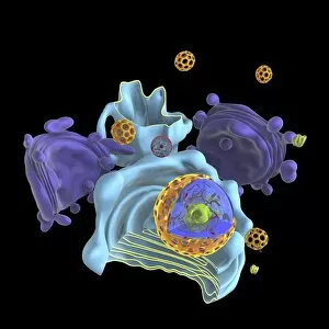
Background imageDNA Collection: Animal cell organelles, artwork C016 / 0619

Animal cell organelles, artwork C016 / 0619
Computer artwork showing the organelles in a eukaryotic cell. This is an animal cell. Structures include the nucleus (centre, red) which has a membrane with nuclear pores

Background imageDNA Collection: Animal cell organelles, artwork C016 / 0621

Animal cell organelles, artwork C016 / 0621
Computer artwork showing the organelles in a eukaryotic cell. This is an animal cell. Structures include the nucleus (centre, red) which has a membrane with nuclear pores

Background imageDNA Collection: Animal cell organelles, artwork C016 / 0620

Animal cell organelles, artwork C016 / 0620
Computer artwork showing the organelles in a eukaryotic cell. This is an animal cell. Structures include the nucleus (centre, orange) which has a membrane with nuclear pores

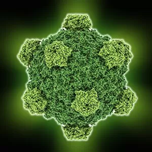
Background imageDNA Collection: Alpha 3 bacteriophage capsid

Alpha 3 bacteriophage capsid, molecular model. Bacteriophages are viruses that infect bacteria, with the capsid forming the bacteriophage head. A capsid has subunits called capsomeres

Background imageDNA Collection: Animal cell organelles, artwork C016 / 0617

Animal cell organelles, artwork C016 / 0617
Computer artwork showing the organelles in a eukaryotic cell. This is an animal cell. Structures include the nucleus (centre, red) which has a membrane with nuclear pores

Background imageDNA Collection: Animal cell organelles, artwork C016 / 0618

Animal cell organelles, artwork C016 / 0618
Computer artwork showing the organelles in a eukaryotic cell. This is an animal cell. Structures include the nucleus (centre, orange) which has a membrane with nuclear pores

Background imageDNA Collection: Animal cell organelles, artwork C016 / 0615

Animal cell organelles, artwork C016 / 0615
Computer artwork showing the organelles in a eukaryotic cell. This is an animal cell. Structures include the nucleus (centre, orange) which has a membrane with nuclear pores

Background imageDNA Collection: Animal cell organelles, artwork C016 / 0616

Animal cell organelles, artwork C016 / 0616
Computer artwork showing the organelles in a eukaryotic cell. This is an animal cell. Structures include the nucleus (centre, orange) which has a membrane with nuclear pores

Background imageDNA Collection: Animal cell organelles, artwork C016 / 0611

Animal cell organelles, artwork C016 / 0611
Computer artwork showing the organelles in a eukaryotic cell. This is an animal cell. Structures include the nucleus (centre, red) which has a membrane with nuclear pores

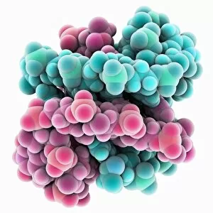
Background imageDNA Collection: DNA hybrid duplex, molecular model

DNA hybrid duplex, molecular model. This model shows a chimeric junction, where a DNA (deoxyribonucleic acid) strand changes from one form to another

Background imageDNA Collection: Animal cell organelles, artwork C016 / 0612

Animal cell organelles, artwork C016 / 0612
Computer artwork showing the organelles in a eukaryotic cell. This is an animal cell. Structures include the nucleus (centre, orange) which has a membrane with nuclear pores

Background imageDNA Collection: Animal cell organelles, artwork C016 / 0605

Animal cell organelles, artwork C016 / 0605
Computer artwork showing the organelles in a eukaryotic cell. This is an animal cell. Structures include the nucleus (centre, red) which has a membrane with nuclear pores

Background imageDNA Collection: Alpha 3 bacteriophage capsid

Alpha 3 bacteriophage capsid, molecular model. Bacteriophages are viruses that infect bacteria, with the capsid forming the bacteriophage head. A capsid has subunits called capsomeres

Background imageDNA Collection: Animal cell organelles, artwork C016 / 0610

Animal cell organelles, artwork C016 / 0610
Computer artwork showing the organelles in a eukaryotic cell. This is an animal cell. Structures include the nucleus (centre, orange) which has a membrane with nuclear pores

Background imageDNA Collection: Animal cell organelles, artwork C016 / 0604

Animal cell organelles, artwork C016 / 0604
Computer artwork showing the organelles in a eukaryotic cell. This is an animal cell. Structures include the nucleus (centre, red) which has a membrane with nuclear pores

Background imageDNA Collection: Animal cell organelles, artwork C016 / 0606

Animal cell organelles, artwork C016 / 0606
Computer artwork showing the organelles in a eukaryotic cell. This is an animal cell. Structures include the nucleus (centre, orange) which has a membrane with nuclear pores

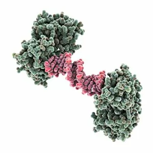
Background imageDNA Collection: Transcription repressor protein and DNA C015 / 5810

Transcription repressor protein and DNA C015 / 5810
Transcription repressor protein and DNA, molecular model. The repressor protein (green) is binding to a strand of DNA (deoxyribonucleic acid, pink and purple)

Background imageDNA Collection: Heat shock transcription factor and DNA C015 / 5558

Heat shock transcription factor and DNA C015 / 5558
Heat shock transcription factor and DNA. Molecular model of the binding domain region (purple) of a heat shock protein transcription factor bound to DNA (pink, deoxyribonucleic acid)

Background imageDNA Collection: Z-DNA tetramer molecule C015 / 6558

Z-DNA tetramer molecule C015 / 6558
Z-DNA (deoxyribonucleic acid) tetramer, molecular model. DNA is composed of two strands twisted into a double helix. This is a tetramer of the molecule, containing four strands

Background imageDNA Collection: SMAD4 protein domain bound to DNA C015 / 6552

SMAD4 protein domain bound to DNA C015 / 6552
SMAD4 protein domain bound to DNA, molecular model. This strand of DNA (deoxyribonucleic acid, green and pink) is surrounded by MH1 domains of the SMAD4 (Mothers against decapentaplegic homolog 4)

Background imageDNA Collection: SMAD4 protein domain bound to DNA C015 / 6551

SMAD4 protein domain bound to DNA C015 / 6551
SMAD4 protein domain bound to DNA, molecular model. This strand of DNA (deoxyribonucleic acid, red and blue) is surrounded by MH1 domains of the SMAD4 (Mothers against decapentaplegic homolog 4)

Background imageDNA Collection: HIV-1 polypurine tract, molecular model C015 / 5822

HIV-1 polypurine tract, molecular model C015 / 5822
HIV-1 polypurine tract. Molecular model of the polypurine tract (PPT, green) from human immunodeficiency virus-1 (HIV-1) complexed with reverse transcriptase (red)

Background imageDNA Collection: HIV-1 polypurine tract, molecular model C015 / 5821

HIV-1 polypurine tract, molecular model C015 / 5821
HIV-1 polypurine tract. Molecular model of the polypurine tract (PPT, red) from human immunodeficiency virus-1 (HIV-1) complexed with reverse transcriptase (green)

Background imageDNA Collection: Transcription repressor protein and DNA C015 / 5811

Transcription repressor protein and DNA C015 / 5811
Transcription repressor protein and DNA, molecular model. The repressor protein (purple) is binding to a strand of DNA (deoxyribonucleic acid, yellow and pink)

Background imageDNA Collection: Female anatomy, artwork

Female anatomy, artwork
Bacteriophage. Artwork of the anatomical structure of a bacteriophage, a virus that targets bacteria. The head (top) contains the DNA (red stands) within the capsid (blue hexagon)

Background imageDNA Collection: DNA MassARRAY analysis C015 / 6522

DNA MassARRAY analysis C015 / 6522
DNA MassARRAY analysis. Close-up of a Sequenom DNA MassARRAY machine. MassARRAY platforms are used for SNP (single-nucleotide polymorphism) genotyping

Background imageDNA Collection: DNA MassARRAY analysis C015 / 6520

DNA MassARRAY analysis C015 / 6520
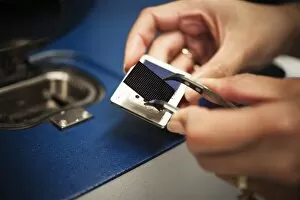
DNA MassARRAY analysis. Technician holding a chip from a Sequenom DNA MassARRAY machine. MassARRAY platforms are used for SNP (single-nucleotide polymorphism) genotyping

Background imageDNA Collection: DNA MassARRAY analysis C015 / 6521

DNA MassARRAY analysis C015 / 6521
DNA MassARRAY analysis. Close-up of a Sequenom DNA MassARRAY machine. MassARRAY platforms are used for SNP (single-nucleotide polymorphism) genotyping

Background imageDNA Collection: DNA MassARRAY analysis C015 / 6518

DNA MassARRAY analysis C015 / 6518
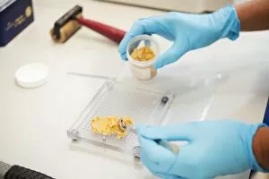
DNA MassARRAY analysis. Technician filling sample plates with resin in a molecular epidemiology lab before running it through a Sequenom DNA MassARRAY machine

Background imageDNA Collection: DNA MassARRAY analysis C015 / 6519

DNA MassARRAY analysis C015 / 6519
DNA MassARRAY analysis. Technician holding chips from a Sequenom DNA MassARRAY machine. MassARRAY platforms are used for SNP (single-nucleotide polymorphism) genotyping

Background imageDNA Collection: DNA MassARRAY analysis C015 / 6517

DNA MassARRAY analysis C015 / 6517
DNA MassARRAY analysis. Technician filling sample plates with resin in a molecular epidemiology lab before running it through a Sequenom DNA MassARRAY machine

Background imageDNA Collection: Androgen receptor, molecular model C015 / 6113

Androgen receptor, molecular model C015 / 6113
Androgen receptor. Molecular model of the DNA-binding region of an androgen receptor (purple and red) complexed with DNA (deoxyribonucleic acid, blue and orange)

Background imageDNA Collection: Fibroblast cell, fluorescent micrograph

Fibroblast cell, fluorescent micrograph
Fibroblast cell. Confocal laser scanning micrograph of a fibroblast cell. The cell nucleus, which contains the cells genetic information, is yellow

Background imageDNA Collection: Androgen receptor, molecular model C015 / 6112

Androgen receptor, molecular model C015 / 6112
Androgen receptor. Molecular model of the DNA-binding region of an androgen receptor (purple and brown) complexed with DNA (deoxyribonucleic acid, turquoise and red)

Background imageDNA Collection: Fibroblast cells, fluorescent micrograph

Fibroblast cells, fluorescent micrograph
Fibroblast cells. Confocal laser scanning micrograph of dividing fibroblast cells. The cell nuclei, which contains the cells genetic information, are yellow

Background imageDNA Collection: Fibroblast cell, fluorescent micrograph

Fibroblast cell, fluorescent micrograph
Fibroblast cell. Confocal laser scanning micrograph of a fibroblast cell. The cell nucleus, which contains the cells genetic information, is blue

Background imageDNA Collection: Fibroblast cell, fluorescent micrograph

Fibroblast cell, fluorescent micrograph
Fibroblast cell. Confocal laser scanning micrograph of a fibroblast cell. The cell nucleus, which contains the cells genetic information, is yellow


All Professionally Made to Order for Quick Shipping


-

Why Choose Us?

We are a leading provider of Art Prints and Photo Gifts since 2004, working in partnership with a range of Sporting Clubs, Charities, Museums and Picture Libraries. A large share of profits from any of their images will go directly towards supporting that charity or club. Our archive of images is carefully curated to bring you a wide range of subjects, including landscapes, wildlife, architecture, and more. We ship from our partner labs in the UK, USA, EU (Netherlands) and Australia.
+

How do I place an order?

Ordering is quick & easy - Just follow 5 Simple Steps:
  1. Find your image: Use our search box or browse our online photo Collections to find the image you want.

  2. Choose your print format: Select your desired product and add it to your cart.

  3. Enter your details: If you're a returning customer, simply enter your email address and password, and we'll fill in your billing and shipping address details. All personal details are held securely and are fully GDPR compliant. As standard, we remove all Personally Identifiable Information after 12 months.

  4. Pay for your purchase: We use state-of-the-art security for online shopping and do not have access to your card details.

  5. Sit back and relax: We'll email you confirmation of your order and when it's dispatched. Registered customers can also track orders in the 'My Account' area.

+

How do I pay for an order?

You can pay for your order with most of the major credit and debit cards, or PayPal. For added security, major financial institutions process payment details separately and securely on our behalf. We do not have access to your online payment card details. Online payments are preferred however we do take phone orders. For UK customers only we accept cheques issued against a UK bank.
+

Is my data safe?

We take data security very seriously. We do not have access to your full card details and all payments are requested over a fully secure connection. Additionally, we fully comply with current European and GDPR legislation, and automatically remove all personal data after at most 12 months (unless you have an account with us and have signed in recently). We also have a strict opt-in policy and would never sell your personal details. Your data is only used to fulfill your order promptly and efficiently.
+

Is wall art ready to hang?

For quick and easy installation all wall art, including framed prints, canvas prints and metal prints are supplied with a ready to hang solution on the back. Generally, saw tooth hangers are applied as they allow wall art to hang flush against the wall. The serrated edge of the hanger prevents the frame from shifting or tilting when hung.
+

Are the photo prints fade resistant?

Yes, we use archival quality photo paper photographic paper for vivid reproduction Prints are an accurate representations of the original artwork, which is preserved for artistic character and authenticity. We guarantee they match previews shown on our web site