Glass Frame > Arts > Artists > C > Thomas Cross
Glass Frame : Cardiac muscle, TEM


![]()
Mounted Prints from Science Photo Library
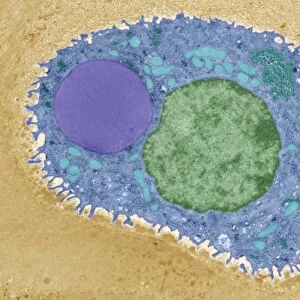
Cardiac muscle, TEM
Cardiac muscle. Coloured transmission electron micrograph (TEM) of cardiac muscle fibrils (orange)from a healthy heart. Mitochondria (pink) supply the muscle cells with energy. The muscle fibrils, or myofibrils, are crossed by transverse tubules (dark orange lines). These tubules mark the division of the myofibrils into contractile units (sarcomeres). Cardiac muscle is under subconscious control and continuously contracts to pump blood around the body without tiring. Magnification: 3600x when printed 10 centimetres across
Science Photo Library features Science and Medical images including photos and illustrations
Media ID 6448627
© THOMAS DEERINCK, NCMIR/SCIENCE PHOTO LIBRARY
Cardiac Cardiology Continuous Contractile Unit Fibre Fibril Fibrils Histological Histology Micrograph Microscope Mitochondrion Muscles Myocardium Myofibril Myofibrils Organelle Organelles Pump Sarcomere Sarcomeres Transmission Electron Transmission Electron Micrograph Transverse Tubules Tubule Units Z Lines False Coloured
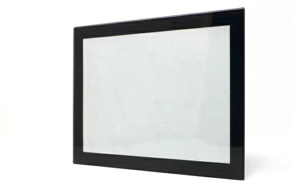
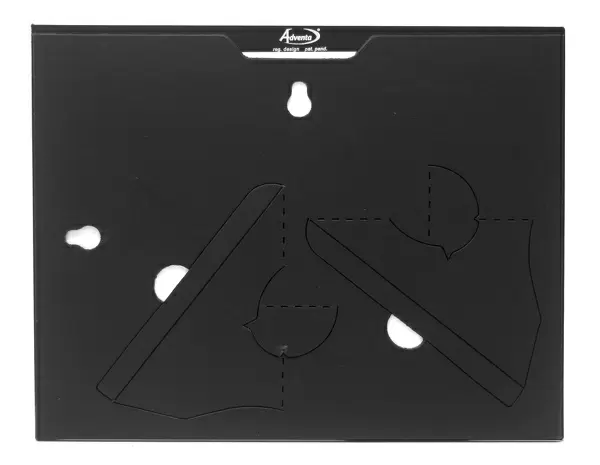
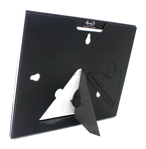
8"x6" Glass Mount
Wall mounted or free-standing, these black edged glass frames feature a smooth chamfered edge and a stylish black border (on back face of the glass). Manufactured from 4mm thick glass, Glass Mounts are a durable, professional way of displaying and protecting your prints. Your 8x6 print is slotted into the back of the frame so can easily be changed if needed.
Tempered Glass Mounts are ideal for wall display, plus the smaller sizes can also be used free-standing via an integral stand
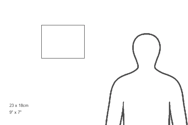
Estimated Image Size (if not cropped) is 20.3cm x 15.2cm (8" x 6")
Estimated Product Size is 22.8cm x 17.7cm (9" x 7")
These are individually made so all sizes are approximate
Artwork printed orientated as per the preview above, with landscape (horizontal) orientation to match the source image.
EDITORS COMMENTS
This vibrant print showcases the intricate beauty of cardiac muscle, a vital component of a healthy heart. In this coloured transmission electron micrograph (TEM), we can observe the mesmerizing orange fibrils that make up the cardiac muscle. These fibrils are responsible for the continuous contraction and relaxation of the heart, allowing it to pump blood throughout our bodies tirelessly. The pink structures within the image represent mitochondria, which play a crucial role in supplying energy to these hardworking muscle cells. Additionally, dark orange lines traverse through the myofibrils, marking transverse tubules that divide them into contractile units called sarcomeres. This division allows for efficient coordination and synchronization during each heartbeat. Cardiac muscle operates under subconscious control, ensuring that our hearts beat rhythmically without us having to consciously think about it. It is fascinating to witness how this biological marvel functions seamlessly day in and day out. With a magnification level of 3600x when printed at 10 centimetres across, this print from Science Photo Library offers an awe-inspiring glimpse into the microscopic world of cardiology and anatomy. Its false-coloured presentation adds depth and clarity to highlight key features such as organelles like mitochondria and structures like z lines. Overall, this image serves as a testament to both the complexity and resilience of our cardiovascular system – an intricate network designed for one purpose: keeping us alive by continuously pumping oxygen-rich blood throughout our bodies.
MADE IN THE UK
Safe Shipping with 30 Day Money Back Guarantee
FREE PERSONALISATION*
We are proud to offer a range of customisation features including Personalised Captions, Color Filters and Picture Zoom Tools
SECURE PAYMENTS
We happily accept a wide range of payment options so you can pay for the things you need in the way that is most convenient for you
* Options may vary by product and licensing agreement. Zoomed Pictures can be adjusted in the Basket.