
Z Lines Collection
"Exploring the Intricacies of Z Lines: A Glimpse into Cardiac Muscle Structure" Z lines, also known as Z discs


All Professionally Made to Order for Quick Shipping
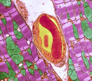
"Exploring the Intricacies of Z Lines: A Glimpse into Cardiac Muscle Structure" Z lines, also known as Z discs, are fascinating structures found within cardiac muscle cells that play a crucial role in muscle contraction. Through advanced imaging techniques such as Transmission Electron Microscopy (TEM) and Scanning Electron Microscopy (SEM), scientists have been able to unravel the intricate details of these essential components. In TEM images, we observe the highly organized arrangement within cardiac muscle fibers. These thin dark bands act as anchors for actin filaments, providing structural support and facilitating efficient contraction. The precise alignment ensures synchronized contractions throughout the heart. Furthermore, SEM reveals an even closer look at the relationship between cardiac muscle and capillaries. Capillaries intimately intertwine with cardiac muscle fibers, supplying oxygen-rich blood necessary for sustained contractions. TEM images showcase how Z lines interact with capillaries on a microscopic level, highlighting their vital role in maintaining proper blood flow. The detailed examination through SEM allows us to appreciate the three-dimensional structure within cardiac muscles. We can observe their periodic pattern along myofibrils and witness their connection to adjacent sarcomeres—a testament to their importance in coordinating muscular movements during each heartbeat. Moreover, SEM provides insights into variations among different regions of cardiac tissue. In some areas captured by SEM imagery, irregularities or disruptions in Z line patterns may be observed—indicating potential abnormalities or disease conditions that warrant further investigation. Overall, these remarkable imaging techniques shed light on the significance in maintaining healthy cardiac function. By understanding their structure at both micro- and nano-levels using TEM and SEM respectively, researchers can gain valuable insights into various cardiovascular disorders while paving the way for innovative therapeutic interventions targeting these fundamental components. Through ongoing research efforts utilizing cutting-edge imaging technologies like TEM and SEM combined with meticulous analysis, we continue to deepen our understanding and their intricate role in cardiac muscle physiology.