Fraud Blocker Skip to main content

Xylem Collection (page 4)

Xylem, the intricate network of plant tissues responsible for water transport and structural support, is a fascinating subject to explore under various microscopes

Background imageXylem Collection: Bamboo stem, light micrograph

Bamboo stem, light micrograph
Bamboo stem. Polarised light micrograph of a section through a bamboo stem (Bambusa vulgaris). Vascular bundles can be seen (yellow/green) containing xylem (larger openings)

Background imageXylem Collection: Maize stem, light micrograph

Maize stem, light micrograph
Maize stem. Polarised light micrograph of a section through a maize plant stem (Zea mays). A vascular bundles (brown) can be seen containing xylem (larger openings)

Background imageXylem Collection: Spiny rush stem, light micrograph

Spiny rush stem, light micrograph
Spiny rush stem. Polarised light micrograph of a section through a spiny rush stem (Juncus actus). Vascular bundles can be seen (purple/red) containing xylem (larger openings)

Background imageXylem Collection: Wheat stem, light micrograph

Wheat stem, light micrograph
Wheat stem. Light micrograph of a section through the stem of a wheat grass (Triticum aestivium). The circular structures (orange and green) are vascular bundles

Background imageXylem Collection: Sugar cane stem, light micrograph

Sugar cane stem, light micrograph
Sugar cane stem. Polarised light micrograph of a section through a sugar cane stem (Saccharum officinareum). Vascular bundles (round blue/purple structures)

Background imageXylem Collection: Reed canary grass stem, light micrograph

Reed canary grass stem, light micrograph
Reed canary grass stem. Light micrograph of a section through a reed canary grass stem (Phalaris arundinacea). Vascular bundles (oval yellow and red structures)

Background imageXylem Collection: Cotton plant root, light micrograph

Cotton plant root, light micrograph
Cotton plant root. Light micrograph of a transverse section through a root of the cotton plant (Gossypium hirsutum). The layers from outer to centre (some very thin)

Background imageXylem Collection: Dragon tree stem, light micrograph

Dragon tree stem, light micrograph
Dragon tree stem. Light micrograph of a section through a dragon tree stem (Dracaena draco). Vascular bundles (round dark structures) can be seen containing xylem (larger openings)

Background imageXylem Collection: Peanut plant stem, light micrograph

Peanut plant stem, light micrograph
Peanut plant stem. Light micrograph of a transverse section through a stem of the peanut plant (Arachis hypogaea). Below the stems outer layer (epidermis) is a cortex of parenchyma cells (blue)

Background imageXylem Collection: Sarsaparilla plant stem, light micrograph

Sarsaparilla plant stem, light micrograph
Maize root. Polarised light micrograph of a section through the root of a sarsaparilla plant (Smilax regelii) showing a vascular cylinder

Background imageXylem Collection: Stinging nettle stem, light micrograph

Stinging nettle stem, light micrograph
Stinging nettle stem. Polarised light micrograph of a transverse section through a stem of the stinging nettle plant (Urtica dioica)

Background imageXylem Collection: Tea leaf, light micrograph

Tea leaf, light micrograph
Tea leaf. Light micrograph of a cross-section through a tea (Camellia sinensis) leaf. The upper and lower epidermis on the surfaces of the leaf are blue

Background imageXylem Collection: Kidney bean stem, light micrograph

Kidney bean stem, light micrograph
Kidney bean stem. Light micrograph of a section through the stem of a kidney bean (Phaseolus vulgaris) plant. The outer layer is the cuticle (brown), with a cortex of parenchyma (yellow) beneath it

Background imageXylem Collection: Broad bean root, light micrograph

Broad bean root, light micrograph
Broad bean root. Light micrograph of a transverse section through the young root of a broad bean (Vicia faba) plant. The outer epidermis is covered in root hairs

Background imageXylem Collection: Kidney bean root, light micrograph

Kidney bean root, light micrograph
Kidney bean root. Light micrograph of a section through the root of a kidney bean (Phaseolus vulgaris) plant. At centre is the pith (blue)

Background imageXylem Collection: Common broom stem, light micrograph

Common broom stem, light micrograph
Common broom stem. Light micrograph of a transverse section through the stem of a common broom (Salicornia europaea) plant

Background imageXylem Collection: Oleander stem, light micrograph

Oleander stem, light micrograph
Oleander stem. Light micrograph of a cross-section through the stem of an oleander (Nerium oleander) tree. The outer layer is a thick cuticle (yellow) and then cork cambium (blue-green)

Background imageXylem Collection: Young pine tree stem, light micrograph

Young pine tree stem, light micrograph
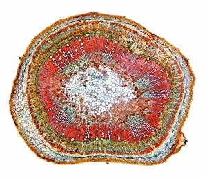
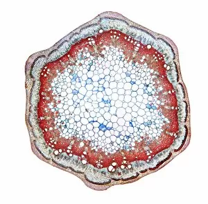
Young pine tree stem. Light micrograph of a transverse section through a two and a half-year-old stem of a pine tree (Pinus sp.)

Background imageXylem Collection: Black burdock stem, light micrograph

Black burdock stem, light micrograph
Black burdock stem. Polarised light micrograph of a transverse section through the stem of a black burdock (Arctium lappa) plant

Background imageXylem Collection: Goosefoot root, light micrograph

Goosefoot root, light micrograph
Goosefoot root. Light micrograph of a section through the root of a goosefoot (Cheopodium sp.) plant. Xylem cells, which transport water around the plant are red

Background imageXylem Collection: Dyers greenweed stem, light micrograph

Dyers greenweed stem, light micrograph
Dyers greenweed stem. Polarised light micrograph of a transverse section through the stem of a dyers greenweed (Genista tinctoria) plant

Background imageXylem Collection: Marsh samphire stem, light micrograph

Marsh samphire stem, light micrograph
Marsh samphire stem. Light micrograph of a transverse section through the stem of a marsh samphire, (Salicornia europaea) plant. This is a succulent, xerophytic, halophyte plant

Background imageXylem Collection: Ginkgo leaf stalk, light micrograph

Ginkgo leaf stalk, light micrograph
Ginkgo leaf stalk. Light micrograph of a transverse section through the leaf stalk (petiole) of a ginkgo tree (Ginkgo biloba)

Background imageXylem Collection: Pine wood structure, light micrograph

Pine wood structure, light micrograph
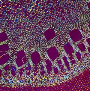
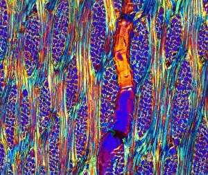
Pine wood structure. Polarised light micrograph of a longitudinal radial section through the stem wood (xylem) of the deal pine tree (Pinus mitis)

Background imageXylem Collection: Begonia stem, light mictograph

Begonia stem, light mictograph
Begoinia stem. Light micrograph of a transverse section through the stem of a begonia (Begonia sp.) plant. At centre is a large area of pith, which contains starch grains (orange) a store of energy

Background imageXylem Collection: Blackcurrant plant stem, light micrograph

Blackcurrant plant stem, light micrograph
Blackcurrant plant stem. Light micrograph of a cross-section through the stem of a blackcurrant (Ribes nigrum) plant. The outer cortex (blue and pale brown) is made of cork cambium tissue

Background imageXylem Collection: Sunflower root, light micrograph

Sunflower root, light micrograph
Sunflower root. Polarised light micrograph of a transverse section through the root of a sunflower (Helianthus annuus) plant

Background imageXylem Collection: Spikenard stem, light micrograph

Spikenard stem, light micrograph
Spikenard stem. Light micrograph of a cross-section through the stem of a Spikenard (Aralia sp.) tree. The dark red cells just beneath the surface bark are collenchyma cells

Background imageXylem Collection: Buttercup root, light micrograph

Buttercup root, light micrograph
Buttercup root. Light micrograph of a transverse section through the root of a buttercup (Ranunculus sp.) plant. At centre is the vascular bundle, comprising xylem (red) and phloem (blue) tissues

Background imageXylem Collection: Rose stem, light micrograph

Rose stem, light micrograph
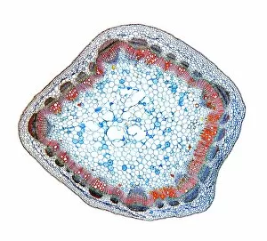
Rose stem. Light micrograph of a cross-section through the stem of a rose (Rosa sp.). The three triangular protrusions are thorns, which protect the plant from being eaten by animals

Background imageXylem Collection: Eucalyptus leaf, light micrograph

Eucalyptus leaf, light micrograph
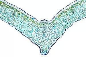
Eucalyptus leaf. Light micrograph of a cross-section through the midrib of a eucalyptus (Eucalyptus globulus) leaf. This xerophytic (arid-adapted)

Background imageXylem Collection: Cedar tree stem, light micrograph

Cedar tree stem, light micrograph
Cedar tree stem. Light micrograph of a transverse section through a stem of a cedar tree (Thujopsis dolobrata). The four ridges on the outer surface are microphyllous leaves

Background imageXylem Collection: Squash root, light micrograph

Squash root, light micrograph
Squash root. Light micrograph of a transverse section through a squash (Cucubita sp.) root. The large vessels (red) are xylem cells, which transport water. Parenchyma tissue is blue

Background imageXylem Collection: Xylem and phloem plant tissue, SEM

Xylem and phloem plant tissue, SEM
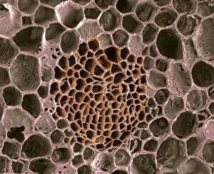
Xylem and phloem plant tissue. Coloured scanning electron micrograph (SEM) of a transverse section through the stem of a woody plant

Background imageXylem Collection: Plant parasite, light micrograph

Plant parasite, light micrograph
Plant parasite. Polarised light micrograph of a transverse section through a plant stem (left) entwined by the common dodder (Cuscuta sp.) parasitic plant (round, right)

Background imageXylem Collection: Dog rose stem, light micrograph

Dog rose stem, light micrograph
Dog rose stem. Polarised light micrograph of a cross-section through the stem of a dog rose (Rosa canina) plant. The outer cortex (black and red) is made of cork cambium tissue

Background imageXylem Collection: Japanese sago palm leaf, light micrograph

Japanese sago palm leaf, light micrograph
Japanese sago palm leaf. Light micrograph of a transverse section through the leaf (pinna) of a Japanese sago palm, cycad, (Cycas revoluta)

Background imageXylem Collection: Sunflower leaf, light micrograph

Sunflower leaf, light micrograph
Sunflower leaf. Light micrograph of a transverse section through the midrib of a sunflower (Helianthus annuus) leaf. The upper and lower epidermis on the surfaces of the leaf are blue

Background imageXylem Collection: Eucalyptus stem, light micrograph

Eucalyptus stem, light micrograph
Eucalyptus stem. Light micrograph of a transverse section through a one-year-old stem of a Eucalyptus (Eucalyptus globulus) plant

Background imageXylem Collection: Mahogany wood structure, light micrograph

Mahogany wood structure, light micrograph
Mahogany wood structure. Polarised light micrograph of a longitudinal tangential section through a woody stem (xylem) of a mahogany (Swietenia sp.) tree

Background imageXylem Collection: Black pepper stem, light micrograph

Black pepper stem, light micrograph
Black pepper stem. Polarised light micrograph of a transverse section through the stem of a black pepper (Pipa nigrum) plant

Background imageXylem Collection: Ginger leaf, light micrograph

Ginger leaf, light micrograph
Ginger leaf. Light micrograph of a transverse section through the midrib of a ginger (Zingiber officinale) leaf. The lower and upper epidermis (blue)

Background imageXylem Collection: Yew tree leaf, light micrograph

Yew tree leaf, light micrograph
Yew tree leaf. Light micrograph of a transverse section through the leaf (pinna) of a yew tree, (Taxus baccata). The structure has xerophytic (drought plants) characteristics

Background imageXylem Collection: Japanese sago palm root, light micrograph

Japanese sago palm root, light micrograph
Japanese sago palm root. Light micrograph of a transverse section through a root of a Japanese sago palm, (Cycas revoluta)

Background imageXylem Collection: Young yew tree stem, light micrograph

Young yew tree stem, light micrograph
Young yew tree stem. Light micrograph of a transverse section through a one-year-old stem of a yew tree (Taxus baccata). The epidermis (outer layer)

Background imageXylem Collection: Willow stem, light micrograph

Willow stem, light micrograph
Willow stem. Light micrograph of a cross-section through the stem of a willow (Salix sp.) tree. At centre is the pith (white), surrounded by woody xylem tissue (pink)

Background imageXylem Collection: Dry rot fungus

Dry rot fungus. Dry rot, unlike its name suggests, infects damp timber. The term refers to the growth of either of two fungi

Background imageXylem Collection: Rotten wood, SEM

Rotten wood, SEM
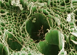
Rotten wood. Coloured scanning electron micrograph (SEM) of a piece of domestic plywood infected with dry rot fungus. The structure of the wood is seen here


All Professionally Made to Order for Quick Shipping

Xylem, the intricate network of plant tissues responsible for water transport and structural support, is a fascinating subject to explore under various microscopes. From scanning electron microscopy (SEM) images capturing the detailed vascular bundles to light micrographs showcasing different plant parts, xylem reveals its beauty and functionality. In a castor oil stem captured by a light micrograph, we witness the complexity tissue as it forms an interconnected system of vessels that efficiently transports water from roots to leaves. Similarly, in a maize root image taken with a light microscope, we can observe the organized arrangement cells that enable efficient uptake of water and nutrients. Moving on to leaf structures, a captivating light micrograph showcases the delicate veins within a water lily leaf. These intricate networks are composed mainly tissue which ensures proper hydration throughout the leaf's surface area. Another stunning SEM image captures silver birch twig's xylem vessels in detail - their elongated shapes perfectly adapted for fluid transportation. Exploring further into plants' underground systems, we encounter an enchanting light micrograph displaying the rhizome structure of a water fern. Xylem tissues play an essential role here too by providing support and facilitating nutrient absorption from surrounding soil particles. Light micrographs also capture xylem wonders in stems like those found in pine trees or pondweeds. Pine tree stems exhibit dense clusters of tracheids – specialized cells within xylem – while pondweed stems showcase long strands forming hollow tubes for efficient water movement. Even at smaller scales observed through SEM imaging techniques, such as examining isolated samples of pure xylem tissue or wood itself under high magnification power; one can appreciate its intricate cellular arrangements that allow for optimal fluid flow and mechanical stability. Lastly, exploring below ground once again brings us to oak roots depicted through another mesmerizing light micrograph.