
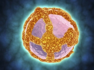
Virus Collection (page 5)
"Unseen Threats: Exploring the Intricacies of Viruses and their Impact on Humanity" Avian flu virus: A microscopic view of the avian flu virus


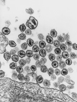


POLIOVIRUS, 1953. An early image of the poliovirus taken by an electron microscope. Photograph, 1953


Digital cross section illustration of ciliate cell showing rhinovirus and antobodies in nasal cavity


All Professionally Made to Order for Quick Shipping
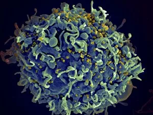
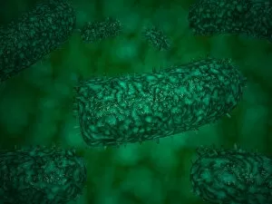
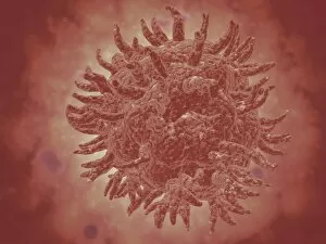
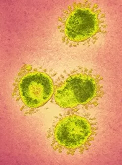
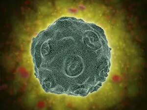
"Unseen Threats: Exploring the Intricacies of Viruses and their Impact on Humanity" Avian flu virus: A microscopic view of the avian flu virus, a notorious pathogen that poses a significant threat to both birds and humans. Unveiling HIV: Delving into the intricate structure of an HIV particle, shedding light on one of the most challenging viruses humanity has faced. Norovirus Particles Revealed: Captured through a powerful TEM microscope, these norovirus particles showcase their unique shape and potential for causing severe gastrointestinal illness. Decoding Hepatitis C: A molecular model displays the complex enzyme responsible for Hepatitis C infection, highlighting ongoing efforts to combat this silent killer. Alexandre Yersin's Discovery: Paying homage to Alexandre Yersin, whose groundbreaking research led to identifying the causative agent behind bubonic plague - Yersinia pestis bacteria. Avian Flu Resurgence: With avian flu outbreaks resurfacing globally, understanding its transmission dynamics becomes crucial in preventing future pandemics. Battling Hepatitis B: Molecular models depict hepatitis B viruses - a major cause of liver disease worldwide - emphasizing the importance of vaccination campaigns and public health initiatives. Empowering Indigenous Communities against Viral Threats in Mexico: Highlighting efforts made towards educating indigenous populations about viral diseases amidst poverty-stricken conditions in Mexico. Coronavirus Artwork Reflects Global Crisis: An artistic representation captures the essence of coronavirus' impact on society, reminding us all to remain vigilant in our fight against this relentless enemy. Broken Tulip Syndrome Exposed: The example of a broken tulip serves as an analogy for how certain plant viruses can devastate entire crops, posing economic challenges for farmers worldwide. Respiratory Syncytial Virus Under Microscope Lens.