Fraud Blocker Skip to main content

Vertebra Collection (page 5)

"Unveiling the Backbone of Life: Exploring the Fascinating World of Vertebrae" A glimpse into history reveals a horse's skull

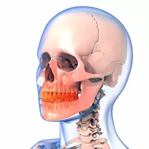
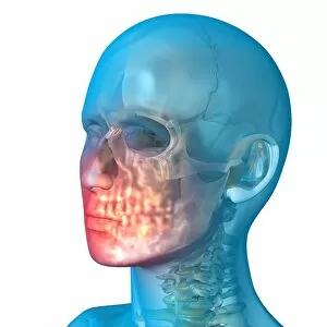
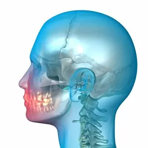
Background imageVertebra Collection: Toothache, artwork C013 / 5927

Toothache, artwork C013 / 5927
Toothache, computer artwork

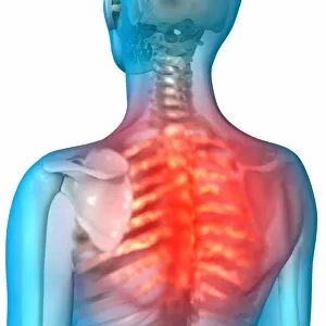
Background imageVertebra Collection: Shoulder pain, artwork C013 / 5925

Shoulder pain, artwork C013 / 5925
Shoulder pain, computer artwork

Background imageVertebra Collection: Toothache, artwork C013 / 5926

Toothache, artwork C013 / 5926
Toothache, computer artwork

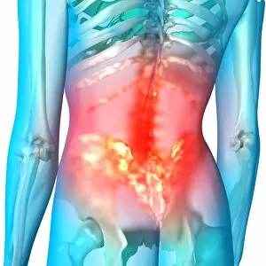
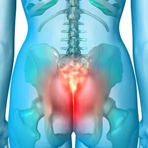
Background imageVertebra Collection: Lower back pain, artwork C013 / 5922

Lower back pain, artwork C013 / 5922
Lower back pain, computer artwork

Background imageVertebra Collection: Neck pain, artwork C013 / 5923

Neck pain, artwork C013 / 5923
Neck pain, computer artwork

Background imageVertebra Collection: Jaw pain, artwork C013 / 5920

Jaw pain, artwork C013 / 5920
Jaw pain, computer artwork

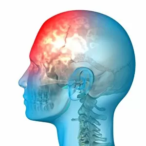
Background imageVertebra Collection: Headache, artwork C013 / 5916

Headache, artwork C013 / 5916
Headache, computer artwork

Background imageVertebra Collection: Headache, artwork C013 / 5915

Headache, artwork C013 / 5915
Headache, computer artwork

Background imageVertebra Collection: Headache, artwork C013 / 5914

Headache, artwork C013 / 5914
Headache, computer artwork

Background imageVertebra Collection: Headache, artwork C013 / 5913

Headache, artwork C013 / 5913
Headache, computer artwork

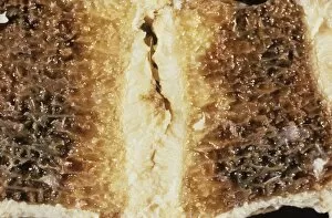
Background imageVertebra Collection: Backbone fat deposition C015 / 6053

Backbone fat deposition C015 / 6053
Backbone fat deposition. Sectioned gross clinical specimen of a vertebra (spinal bone) that displays adiposis (fat deposition, yellow) in the medullary (inner) region

Background imageVertebra Collection: Neck pain, artwork C013 / 8811

Neck pain, artwork C013 / 8811
Neck pain, computer artwork

Background imageVertebra Collection: Lower back pain, artwork C013 / 8810

Lower back pain, artwork C013 / 8810
Lower back pain, computer artwork

Background imageVertebra Collection: Natural history specimens, 18th century C013 / 7818

Natural history specimens, 18th century C013 / 7818
Natural history specimens. 18th-century artwork of various natural history specimens, including vertebrae (top, fish vertebra at left), elephant molars (centre), a diagram of a drumming-well

Background imageVertebra Collection: Back pain, artwork C013 / 7814

Back pain, artwork C013 / 7814
Back pain, computer artwork

Background imageVertebra Collection: Toothache, artwork C013 / 7813

Toothache, artwork C013 / 7813
Toothache, computer artwork

Background imageVertebra Collection: Toothache, artwork C013 / 7812

Toothache, artwork C013 / 7812
Toothache, computer artwork

Background imageVertebra Collection: Shoulder pain, artwork C013 / 7810

Shoulder pain, artwork C013 / 7810
Shoulder pain, computer artwork

Background imageVertebra Collection: Toothache, artwork C013 / 7811

Toothache, artwork C013 / 7811
Toothache, computer artwork

Background imageVertebra Collection: Headache, artwork C013 / 7797

Headache, artwork C013 / 7797
Headache, computer artwork

Background imageVertebra Collection: Headache, artwork C013 / 7796

Headache, artwork C013 / 7796
Headache, computer artwork

Background imageVertebra Collection: Headache, artwork C013 / 7795

Headache, artwork C013 / 7795
Headache, computer artwork

Background imageVertebra Collection: Coccyx pain, artwork C013 / 7793

Coccyx pain, artwork C013 / 7793
Coccyx (tailbone) pain, computer artwork

Background imageVertebra Collection: Neck pain C013 / 6061

Neck pain C013 / 6061
MODEL RELEASED. Neck pain. Young woman holding her painful or stiff neck. Possible causes of neck pain may include strains of the neck muscles, damage to the joints of the neck bones (vertebrae)

Background imageVertebra Collection: Neck pain C013 / 6060

Neck pain C013 / 6060
MODEL RELEASED. Neck pain. Young woman holding her painful or stiff neck. Possible causes of neck pain may include strains of the neck muscles, damage to the joints of the neck bones (vertebrae)

Background imageVertebra Collection: Slipped intervertebral disc, artwork C013 / 4663

Slipped intervertebral disc, artwork C013 / 4663
Slipped intervertebral disc. Computer artwork of a side view of the vertebral column in a case of a slipped (herniated) disc

Background imageVertebra Collection: Myotragus

Myotragus
This animal which lived around 6000 years ago was from the antelope group. Its remains have been found in the Balearic islands

Background imageVertebra Collection: Dinosaur vertebrae

Dinosaur vertebrae
From a Palaeontology field trip in Niger, West Africa

Background imageVertebra Collection: Healthy kidneys, CT scan

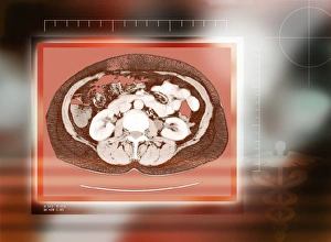
Healthy kidneys, CT scan
Healthy kidneys, coloured computed tomography (CT) scan. The kidneys are the two C-shaped white organs either side of the spinal vertebra (centre). Below the spine and kidneys are back muscles
Background imageVertebra Collection: Kidneys, abdominal CT scan

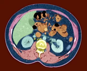
Kidneys, abdominal CT scan
Normal kidneys. Coloured axial (horizontal) CT (computed tomography) scan through a human abdomen at the level of the kidneys. The front of the body is at top
Background imageVertebra Collection: Head and chest anatomy

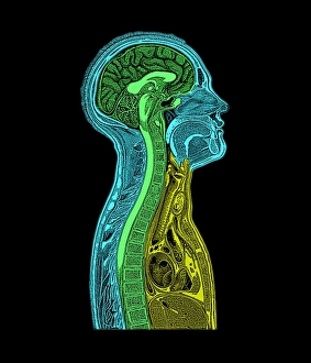
Head and chest anatomy. Historical artwork of a human head and upper torso, cut vertically in half and seen sideways. In the head, layers of skin and bone cover the brain (purple)

Background imageVertebra Collection: Coccyx and lower back, X-ray

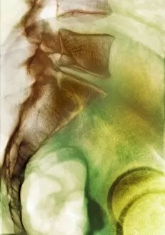
Coccyx and lower back, X-ray
Coccyx and lower back. Coloured X-ray of a side view of the coccyx and lower back of a healthy adult man. The front of the body is at right

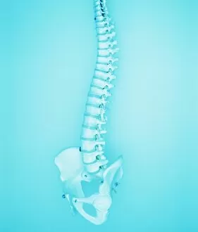
Background imageVertebra Collection: Spine

Spine, computer artwork. Human skeleton with a blue spine. The spine, or backbone, supports the back and protects the spinal cord

Background imageVertebra Collection: Human spine

Human spine. Model of the human backbone or spine. The spine is a flexible column of small compact bones called vertebrae

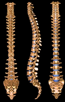
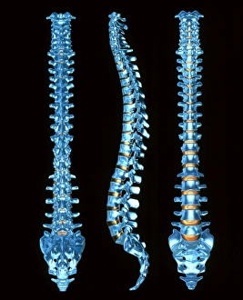
Background imageVertebra Collection: Computer artwork of three views of a human spine

Computer artwork of three views of a human spine
Spine. Computer illustration of three views of a healthy human spine. It is seen from the back at left, the side at centre, and from the front at right

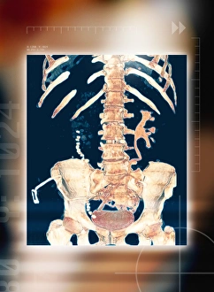
Background imageVertebra Collection: Abdominal bones, 3-D CT scan

Abdominal bones, 3-D CT scan
Abdominal bones, coloured 3-D computer tomography (CT) scan. The lower part of the ribcage is in upper frame, with the pelvis in lower frame. They are connected by the spine, which runs down centre

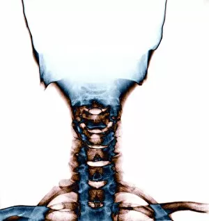
Background imageVertebra Collection: Neck and skull, X-ray

Neck and skull, X-ray
Neck and skull. Coloured X-ray of a normal human neck (lower centre) and part of the skull (upper centre). The neck joins the head to the body

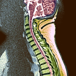
Background imageVertebra Collection: Healthy spine

Healthy spine. Coloured magnetic resonance imaging (MRI) scan of a healthy human spine seen from the side (sagittal side). The front of the spine is on the left

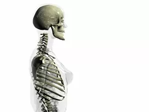
Background imageVertebra Collection: Upper body skeleton

Upper body skeleton. Computer artwork of a glass- effect human female body in side view showing the skeletal structure of the head and torso

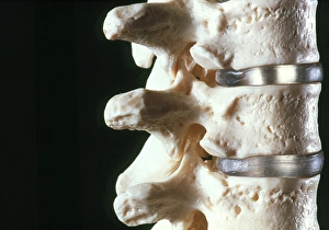
Background imageVertebra Collection: Model of three vertebrae and intervertebral discs

Model of three vertebrae and intervertebral discs
Vertebrae. Model of three vertebrae from the human spine. The vertebrae are separated by intervertebral discs (grey). Vertebrae are bones with a wide

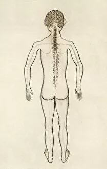
Background imageVertebra Collection: Spinal anatomy, 14th century artwork

Spinal anatomy, 14th century artwork. The spinal cord is shown running down the back, with eighteen spinal nerves braching off through holes in the vertebrae (spinal bones)

Background imageVertebra Collection: Human spine, three views

Human spine, three views
Spine. Computer illustration of three views of a healthy human spine. It is seen from the back at left, the side at centre, and from the front at right

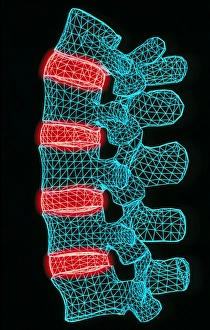
Background imageVertebra Collection: Computer artwork of the five lumbar vertebrae

Computer artwork of the five lumbar vertebrae
Lumbar spine. Computer illustration of the five lumbar vertebrae (blue) of the human spine. The lumbar vertebrae are the largest of the unfused vertebrae in the backbone

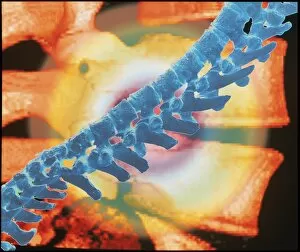
Background imageVertebra Collection: Computer artwork of a spine depicting back pain

Computer artwork of a spine depicting back pain
Back pain. Abstract computer artwork of back pain showing radiating circles coming from a spine (blue) on a background of a close-up of the vertebrae

Background imageVertebra Collection: Artwork of causes of neck pain in cervical spine

Artwork of causes of neck pain in cervical spine
Pain in the neck. Illustration of the cervical vertebrae - the bones of the neck - showing causes of neck pain. A tumour of the spinal cord (red)

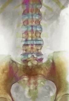
Background imageVertebra Collection: Spondylitis, X-ray

Spondylitis, X-ray
Spondylitis. Coloured X-ray of the lower spine and pelvis of a patient with spondylitis, an inflammation of the synovial joints of the backbone

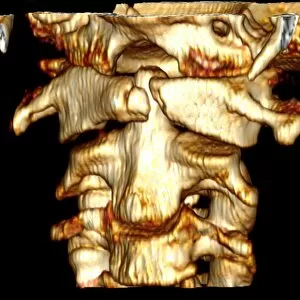
Background imageVertebra Collection: Fractured atlas vertebra, 3D CT scan

Fractured atlas vertebra, 3D CT scan
Fractured atlas vertebra, coloured 3D CT (computed tomography) scan. Posterior view of a fractured atlas vertebra (break at upper centre) of a 34 year old man. Part of the skull can be seen at top

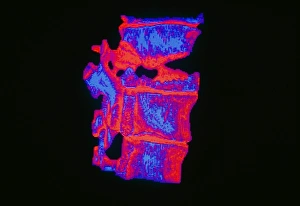
Background imageVertebra Collection: Coloured 3-D CT scan of a fractured vertebra

Coloured 3-D CT scan of a fractured vertebra


All Professionally Made to Order for Quick Shipping

"Unveiling the Backbone of Life: Exploring the Fascinating World of Vertebrae" A glimpse into history reveals a horse's skull, showcasing the intricate vertebrae that form its backbone. Delving deeper, a diagram illustrates the human brain and spinal column, highlighting how vital vertebrae are to our nervous system. With a camera in hand, an individual captures an X-ray image, unveiling the hidden secrets within vertebral structures. Journeying back in time, we encounter a Liopleurodon vertebra—a relic from prehistoric oceans—reminding us of ancient creatures' remarkable skeletal systems. Witnessing neck vertebrae extended through an X-ray image showcases their flexibility and adaptability for various movements. An artistically colored X-ray depicts lumbar vertebrae—the foundation of our spine—revealing their strength and interconnectedness. Examining a normal neck through an X-ray unveils its structural integrity—an essential component for supporting our head and facilitating movement. Immerse yourself in paleontological wonders with a 3D rendering of an Ankylosaurus dinosaur skeleton, emphasizing its robust vertebral framework. Peering inside another 3D rendering—this time featuring a Tyrannosaurus Rex dinosaur skeleton—we marvel at how these extinct giants relied on their powerful spines for survival. Unearthed from centuries past is an illustration depicting the nervous system as understood during the 18th century—a testament to humanity's evolving knowledge about vertebral significance over time. In modern times, workers collaborate to repair damaged spines—an inspiring reminder of medical advancements aimed at restoring mobility and quality of life for individuals facing spinal issues (Spine with workers; spine repair F007 / 9907).