Fraud Blocker Skip to main content

Venous Collection

"Exploring the Intricate Network Pathways

Background imageVenous Collection: Anatomy of human venous return system in the upper torso

Anatomy of human venous return system in the upper torso
Anatomy of human venous return system of venal valves in the upper torso. Lithograph from Lorenz Okens Universal Natural History, Allgemeine Naturgeschichte fur alle Stande, Stuttgart, 1839

Background imageVenous Collection: Vascular System of the Body (engraving) (b / w photo)

Vascular System of the Body (engraving) (b / w photo)
XJF143296 Vascular System of the Body (engraving) (b/w photo) by Vesalius, Andreas (1514-64); Private Collection; Belgian, out of copyright

Background imageVenous Collection: Vascular system of the head, MRI scan

Vascular system of the head, MRI scan
Vascular system of the head. Magnetic resonance imaging (MRI) angiogram of a sagittal section through the head of a healthy 43-year-old, showing the blood vessels (white) of the brain and neck

Background imageVenous Collection: Lung alveoli anatomy, artwork C016 / 7680

Lung alveoli anatomy, artwork C016 / 7680
Lung alveoli anatomy. Artwork showing the structure of the lung alveoli, where gas exchange occurs between the respiratory and circulatory systems

Background imageVenous Collection: Dental filling polymerisation, artwork C014 / 2025

Dental filling polymerisation, artwork C014 / 2025
Dental filling polymerisation. Artwork of a cross-section of a human tooth, with a filling (white) being set using an ultraviolet light lamp tool

Background imageVenous Collection: Thrombosed blood vessel, artwork C013 / 4649

Thrombosed blood vessel, artwork C013 / 4649
Thrombosed blood vessel. Computer artwork of a blood clot (thrombus) formed on the internal wall of a blood vessel

Background imageVenous Collection: Nephron structure, artwork

Nephron structure, artwork
Nephron structure. Artwork showing the structure of a nephron, the functional filtering unit of a kidney. The function of the kidney is to filter waste products such as urea from the blood

Background imageVenous Collection: Woodcut from the third book of Andreas Vesalius De Humani Corporis Fabrica

Woodcut from the third book of Andreas Vesalius De Humani Corporis Fabrica, published in 1543 at Basel
VESALIUS: VENOUS SYSTEM. Woodcut from the third book of Andreas Vesalius De Humani Corporis Fabrica, published in 1543 at Basel

Background imageVenous Collection: Cardiovascular system, artwork F006 / 4620

Cardiovascular system, artwork F006 / 4620
Cardiovascular system, computer artwork

Background imageVenous Collection: Cardiovascular system, artwork F006 / 4611

Cardiovascular system, artwork F006 / 4611
Cardiovascular system, computer artwork

Background imageVenous Collection: Cardiovascular system, artwork F006 / 4600

Cardiovascular system, artwork F006 / 4600
Cardiovascular system, computer artwork

Background imageVenous Collection: Cardiovascular system, artwork F006 / 4596

Cardiovascular system, artwork F006 / 4596
Cardiovascular system, computer artwork

Background imageVenous Collection: Cardiovascular system, artwork F006 / 4602

Cardiovascular system, artwork F006 / 4602
Cardiovascular system, computer artwork

Background imageVenous Collection: Deep abdominal organs, 1880 artwork C017 / 6913

Deep abdominal organs, 1880 artwork C017 / 6913
Deep abdominal organs. Historical anatomical artwork of a dissected abdomen seen from the front, showing the deep-seated organs and major blood vessels after the removal of the intestines

Background imageVenous Collection: Thrombophlebitis in the brain, MRI scans

Thrombophlebitis in the brain, MRI scans
Thrombophlebitis in the brain. Coloured magnetic resonance imaging (MRI) angiograms comparing the blood vessels (white) of the brain and neck in a healthy 43-year-old (left)

Background imageVenous Collection: Thrombophlebitis in the brain, 3D CT scan

Thrombophlebitis in the brain, 3D CT scan
Thrombophlebitis in the brain. Coloured 3D computed tomography (CT) angiogram of the blood vessels (blue) in the brain of a 48-year-old with thrombophlebitis of the superior sagittal sinus

Background imageVenous Collection: Shark heart-gill anatomy, artwork C016 / 6847

Shark heart-gill anatomy, artwork C016 / 6847
Shark heart-gill anatomy. Artwork of the heart and gill anatomy of a shark, seen from the underside, with the head at right (teeth at centre right)

Background imageVenous Collection: Total mesorectal excision, artwork C016 / 6996

Total mesorectal excision, artwork C016 / 6996
Total mesorectal excision (TME). Artwork of the pelvis and the area of the colon and rectum that is removed in total mesorectal excision

Background imageVenous Collection: Dental implant, artwork

Dental implant, artwork
Dental implant. Artworks showing the process of replacing a tooth (incisor) with a dental implant. At upper left, front lower teeth and a dental instrument are shown in a case of receding

Background imageVenous Collection: Lewis-mckenzie polygraph, circa 1900 C017 / 6947

Lewis-mckenzie polygraph, circa 1900 C017 / 6947
Lewis-McKenzie polygraph, made in 1900 by Cambridge and Paul Instrument Co. This device was used to graphically record venous and arterial pulses simultaneously

Background imageVenous Collection: High endothelial venule, TEM C014 / 1446

High endothelial venule, TEM C014 / 1446
High endothelial venule. Transmission electron micrograph (TEM) of a section through a high endothelial venule (HEV). Venules are tiny veins that transport deoxygenated blood from the capillary beds

Background imageVenous Collection: High endothelial venule, TEM C014 / 1445

High endothelial venule, TEM C014 / 1445
High endothelial venule. Transmission electron micrograph (TEM) of a section through a high endothelial venule (HEV). Venules are tiny veins that transport deoxygenated blood from the capillary beds

Background imageVenous Collection: Eye anatomy, artwork C016 / 8742

Eye anatomy, artwork C016 / 8742
Eye anatomy, artwork. The eye is seen in cutaway format from the side. Structures shown include the lens (light blue, lower left), its attachment points, the iris

Background imageVenous Collection: Kidney blood vessels, confocal micrograph C014 / 4610

Kidney blood vessels, confocal micrograph C014 / 4610
Kidney blood vessels. Laser scanning confocal micrograph (LSCM) of a section through a human kidney, showing a blood vessel (vertical, centre)

Background imageVenous Collection: Blood vessels, light micrograph C014 / 4562

Blood vessels, light micrograph C014 / 4562
Blood vessels. Light micrograph of a section through healthy human tissue showing an artery (large, red) and vein (small, red), surrounded by connective tissue (yellow)

Background imageVenous Collection: Blood sampling C015 / 3466

Blood sampling C015 / 3466
Blood sampling. Blood sample being taken from a patients arm. The sample of blood will be labelled and sent for testing. Blood is analysed for levels of components such as cholesterol and glucose

Background imageVenous Collection: Blood sampling C015 / 3465

Blood sampling C015 / 3465
Blood sampling. Blood sample being taken from a patients arm. The sample of blood will be labelled and sent for testing. Blood is analysed for levels of components such as cholesterol and glucose

Background imageVenous Collection: Human eye, infrared image C013 / 7322

Human eye, infrared image C013 / 7322
MODEL RELEASED. Human eye, close-up infrared image. This is the right-hand eye of a 21-year-old man, with raised eyebrow and distended pupil

Background imageVenous Collection: Human arm, infrared image C013 / 7325

Human arm, infrared image C013 / 7325
MODEL RELEASED. Human arm. Infrared image of the anterior surface of the right-hand arm of a 24-year-old man. Infrared lighting can be used to visualize subcutaneous structures

Background imageVenous Collection: Red blood cells, artwork C013 / 4616

Red blood cells, artwork C013 / 4616
Red blood cells. Computer artwork of red blood cells (erythrocytes) flowing through a blood vessel

Background imageVenous Collection: Red blood cells, artwork C013 / 4615

Red blood cells, artwork C013 / 4615
Red blood cells. Computer artwork of red blood cells (erythrocytes) flowing through a blood vessel

Background imageVenous Collection: Venous System 18th C

Venous System 18th C
Veins of the human body Date: Circa 1760

Background imageVenous Collection: Circulation

Circulation, computer artwork. Abstract image of deoxygenated (blue) and oxygenated (red) blood entering the body through the arms and mixing in the abdomen

Background imageVenous Collection: Blood vessel types, artwork

Blood vessel types, artwork
Blood vessel types. Artworks of the three types of blood vessels, from left: arteries, veins, and capillaries. Arteries are thick blood vessels carrying oxygenated blood around the body away from

Background imageVenous Collection: Deep vein thrombosis, artwork

Deep vein thrombosis, artwork
Deep vein thrombosis. Computer artwork showing blood clots in a leg (brown lumps, bottom right), caused by deep vein thrombosis (DVT), moving to blood vessels in the lungs (blue, top right)

Background imageVenous Collection: Normal leg veins, angiogram

Normal leg veins, angiogram
Normal leg veins, coloured angiogram (blood vessel X-ray)

Background imageVenous Collection: Blood vessel structure, artwork

Blood vessel structure, artwork
Blood vessel structure. Artwork showing the layers of tissue in the walls of a vein. In the lumen of the vein are valves (grey) that prevent the back flow of blood

Background imageVenous Collection: Venous valve, artwork

Venous valve, artwork
Venous valve. Artwork showing the anatomy of a vein. Veins carry deoxygenated blood from the body to the heart and lungs. Within hte lumen of the vein are venous valves


All Professionally Made to Order for Quick Shipping

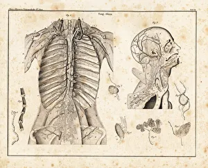
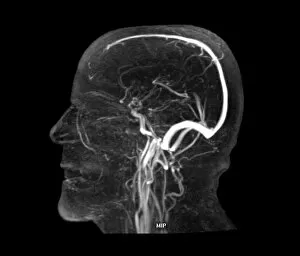
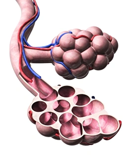
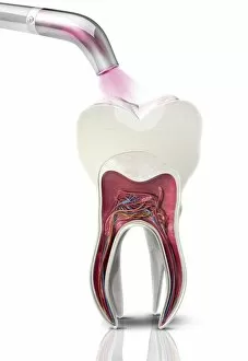
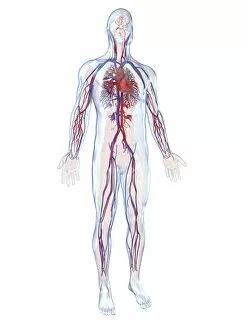
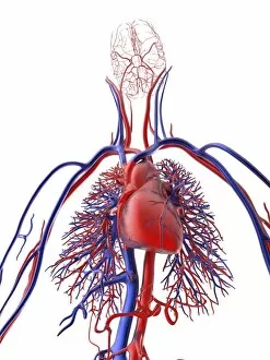
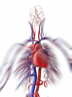
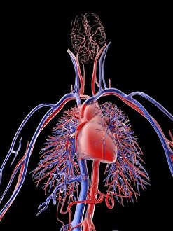
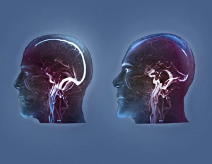
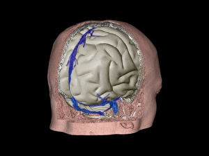
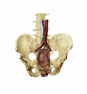
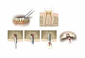
"Exploring the Intricate Network Pathways: From Thyroid Gland Capillaries to Nephron Structures" Delve into the fascinating world anatomy with a captivating array of images and artwork. Witness the intricate web of thyroid gland capillaries, captured in stunning detail through scanning electron microscopy (SEM). Marvel at the complexity of thyroid gland blood vessels, also revealed by SEM, showcasing their vital role in hormone production. Embark on a journey through the cardiovascular system as depicted in exquisite artwork. Transport yourself back to the 16th century with a remarkable illustration depicting heart anatomy, showcasing how our understanding has evolved over centuries. Explore an engraving portraying the vascular system of the body, offering insight into its interconnectedness and importance for overall health. Discover the wonders of modern medical imaging technology as you peer into an MRI scan revealing the vascular system of the head. Gain a deeper appreciation for dental procedures with an artistic representation capturing dental filling polymerization - highlighting how even seemingly unrelated fields rely on proper venous circulation. Witness both beauty and pathology within this vast network. Admire an intricately crafted artwork illustrating thrombosed blood vessels - reminding us that maintaining healthy veins is crucial for optimal well-being. Immerse yourself in another masterpiece from Andreas Vesalius' De Humani Corporis Fabrica, published centuries ago but still revered today for its accurate depiction of human anatomy. From microscopic structures to grand engravings, these visual cues offer glimpses into our complex venous return system spanning across our upper torso and entire body. Appreciate every twist and turn as you navigate through this lifeline that ensures oxygen-rich blood reaches all corners efficiently. Intriguingly intertwined yet uniquely diverse, exploring "venous" reveals not only scientific marvels but also showcases art's ability to capture anatomical wonders throughout history – reminding us once again that knowledge truly knows no boundaries or limitations.