Fraud Blocker Skip to main content

Tumor Collection

DDE-90026224, DDE-90026223, DDE-90026222, DDE-90026221, DDE-90026220, DDE-90026219

Background imageTumor Collection: DDE-90026224

DDE-90026224
Australia, Tasmania. Tasmanian Devil at Trowunna Wildlife Sanctuary. Date: 27/07/2006

Background imageTumor Collection: DDE-90026223

DDE-90026223
Australia, Tasmania. Tasmanian Devil at Trowunna Wildlife Sanctuary. Date: 27/07/2006

Background imageTumor Collection: DDE-90026222

DDE-90026222
Australia, Tasmania. Tasmanian Devil at Trowunna Wildlife Sanctuary. Date: 27/07/2006

Background imageTumor Collection: DDE-90026221

DDE-90026221
Tasmanian Devil at Trowunna Wildlife Sanctuary, Tasmania, Australia Date: 27/07/2006

Background imageTumor Collection: DDE-90026220

DDE-90026220
Australia, Tasmania. Tasmanian Devil at Trowunna Wildlife Sanctuary. Date: 27/07/2006

Background imageTumor Collection: DDE-90026219

DDE-90026219
Australia, Tasmania. Tasmanian Devil at Trowunna Wildlife Sanctuary. Date: 27/07/2006

Background imageTumor Collection: DDE-90026218

DDE-90026218
Australia, Tasmania. Tasmanian Devil at Trowunna Wildlife Sanctuary. Date: 27/07/2006

Background imageTumor Collection: Joseph Carey Merrick, 1889 (b / w photo)

Joseph Carey Merrick, 1889 (b / w photo)
2600202 Joseph Carey Merrick, 1889 (b/w photo) by English Photographer, (19th century); (add.info.: Joseph Carey Merrick (1862-90) English man known as the " Elephant Man"

Background imageTumor Collection: Anterior view female anatomy showing breast tissue with a tumor

Anterior view female anatomy showing breast tissue with a tumor
Anatomy, Diagram, Illustration, Medical, Biology, Surgery, Cancer, See Through, Cut Out, White Background, Vertical, Artwork, Female Likeness, Breast, Color Image, Front View

Background imageTumor Collection: Medical ilustration of a pilonidal cyst near the natal cleft of the buttocks

Medical ilustration of a pilonidal cyst near the natal cleft of the buttocks

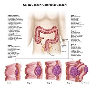
Background imageTumor Collection: Medical illustration depicting the different stages of colon cancer

Medical illustration depicting the different stages of colon cancer

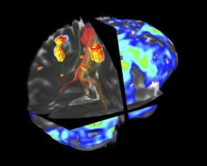
Background imageTumor Collection: Brain tumour, fMRI and tractography C017 / 7102

Brain tumour, fMRI and tractography C017 / 7102
Brain tumour, fMRI and tractography. Combined functional magnetic resonance imaging (fMRI, blue and green) and tractography (yellow and red) imaging of a brain with a tumour (upper left)

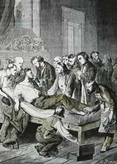
Background imageTumor Collection: MORTON, William Thomas Green (1819-1868). American

MORTON, William Thomas Green (1819-1868). American dentist that considered himself as the inventor and revealer of the use of ether for surgical anesthesia

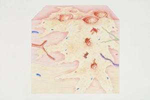
Background imageTumor Collection: Cross-section diagram of a cancerous tumour including calcium deposits, blood vessels

Cross-section diagram of a cancerous tumour including calcium deposits, blood vessels, tumour outgrowth, epithelial layer, ulcerated area, bleeding, nerve fibres, dead tissue and a lymph vessel

Background imageTumor Collection: Kaposis sarcoma on the skin of an AIDS patient

Kaposis sarcoma on the skin of an AIDS patient

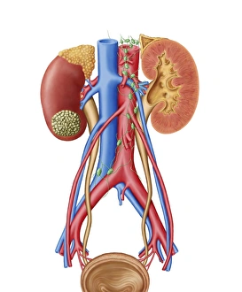
Background imageTumor Collection: Early stage of kidney cancer with a tumor visible on the kidney

Early stage of kidney cancer with a tumor visible on the kidney

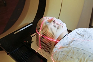
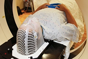
Background imageTumor Collection: Lasers used to position a patient wearing a short face mask in a computed tomography

Lasers used to position a patient wearing a short face mask in a computed tomography
Lasers are used to position a patient model wearing a short face mask in a computed tomography (CT) scanner to obtain images of a tumor before radiation therapy

Background imageTumor Collection: Breast Cancer. Oil Painting C017 / 1273
Breast Cancer. Oil Painting C017 / 1273
Breast Cancer. Painting of a woman after breast removal surgery (mastectomy) Oil on Canvas by Nurit Shany

Background imageTumor Collection: Brain tumour, DTI modelling C017 / 7060

Brain tumour, DTI modelling C017 / 7060
Brain tumour. Computer artwork of nerve pathways (coloured) in a brain with a tumour (white/red, upper right). The brain is seen from the front. Brain tumours can be benign or malignant (cancerous)

Background imageTumor Collection: Brain cancer, DTI and 3D CT scans C016 / 6414

Brain cancer, DTI and 3D CT scans C016 / 6414
Brain cancer. Coloured composite image of diffusion tensor imaging (DTI) scans of the brains, and 3D computed tomography (CT) scans of the skulls, of two 35 year old patients

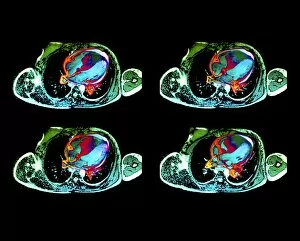
Background imageTumor Collection: Cardiac lymphoma, MRI scans

Cardiac lymphoma, MRI scans
Brain tumour. Coloured magnetic resonance imaging (MRI) scans of axial sections through the chest of a 22-year-old patient, showing a tumorous lesion invading the right ventricle of the heart (large)

Background imageTumor Collection: Uterine fibroids, MRI scan C018 / 0466

Uterine fibroids, MRI scan C018 / 0466
Uterine fibroids. Magnetic resonance imaging (MRI) scan of the pelvis of a 47 year old female with two large fibroids (centre) in her uterus

Background imageTumor Collection: Colon cancer, endoscopic view C016 / 8308

Colon cancer, endoscopic view C016 / 8308
Colon cancer. Endoscopic view of a malignant (cancerous) adenocarcinoma (upper right) in a patients ascending colon (large intestine)

Background imageTumor Collection: Secondary lung cancers, X-ray

Secondary lung cancers, X-ray
Secondary lung cancers. X-ray of the chest of a 60 year old male showing malignant (cancerous) tumours (white) in the lungs

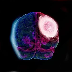
Background imageTumor Collection: Brain tumour, 3D-MRI scan

Brain tumour, 3D-MRI scan
Brain tumour. Coloured three-dimensional model of a male head, made up from numerous magnetic resonance imaging (MRI) scans

Background imageTumor Collection: Cross-section diagram of a non-cancerous tumour including a fibrous capsule

Cross-section diagram of a non-cancerous tumour including a fibrous capsule, tissue layer and blood vessel

Background imageTumor Collection: Digital illustration of angiogenesis process showing dormant tumour invading tissue, growing to mali

Digital illustration of angiogenesis process showing dormant tumour invading tissue, growing to mali

Background imageTumor Collection: Cross section biomedical illustration of breast cancer tumour

Cross section biomedical illustration of breast cancer tumour and lymph nodes requiring mastectomy procedure

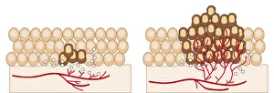
Background imageTumor Collection: Cross section biomedical illustration of cancer cells forming tumour at third doubling stage

Cross section biomedical illustration of cancer cells forming tumour at third doubling stage

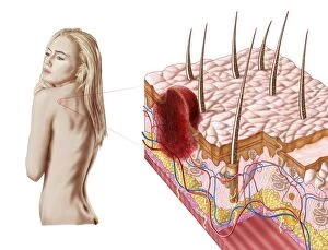
Background imageTumor Collection: Illustration of an atypical growth on the skin that could be a sign of skin cancer

Illustration of an atypical growth on the skin that could be a sign of skin cancer
Illustration of an atypical growth on the skin that could be a warning sign of skin cancer. Sudden change in the appearance, shape, color, size

Background imageTumor Collection: Conceptual image of the Human Papilloma Virus

Conceptual image of the Human Papilloma Virus (HPV). HPV is a virus from the papillomavirus family that affects human skin and the moist membranes that line the body

Background imageTumor Collection: Cancer cell with red blood cell flow

Cancer cell with red blood cell flow

Background imageTumor Collection: Artist depiction of mesothelioma in the lungs / abdominal cavity

Artist depiction of mesothelioma in the lungs / abdominal cavity
Artist depiction of mesothelioma in the lungs/abdominal cavity

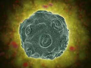
Background imageTumor Collection: Conceptual image of polyomavirus

Conceptual image of polyomavirus

Background imageTumor Collection: Dermal nevi are often elevated about the skin

Dermal nevi are often elevated about the skin and are common on the head and neck area. Coloration may be similar to background skin color as shown here or may be more darkly pigmented

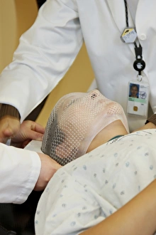
Background imageTumor Collection: Radiation therapists fit a short face mask to a patient during radiation therapy

Radiation therapists fit a short face mask to a patient during radiation therapy
Two radiation therapists fit a short face mask to a patient model to steady the head during radiation therapy

Background imageTumor Collection: Conceptual image of a tumor in human brain

Conceptual image of a tumor in human brain

Background imageTumor Collection: Conceptual image of cancer virus

Conceptual image of cancer virus

Background imageTumor Collection: Microscopic view of a group of macrophages

Microscopic view of a group of macrophages. Macrophages contribute to tumor growth and progression. Attracted to oxygen-starved (hypoxic) and necrotic tumor cells they promote chronic inflammation

Background imageTumor Collection: Patient positioned in computed tomography scanner

Patient positioned in computed tomography scanner
A patient model wearing a short face mask is positioned in a computed tomography (CT) scanner to obtain images of a tumor before radiation therapy

Background imageTumor Collection: Ewings sarcoma locations on the skeleton

Ewings sarcoma locations on the skeleton and detail of tumor on head of femur

Background imageTumor Collection: Microscopic view of cancer virus

Microscopic view of cancer virus

Background imageTumor Collection: Isolated cancer cell Macrophage

Isolated cancer cell Macrophage. Macrophages contribute to tumor growth and progression. Attracted to oxygen-starved (hypoxic) and necrotic tumor cells they promote chronic inflammation

Background imageTumor Collection: Neurofibromatosis, a genetic disorder of the nervous system

Neurofibromatosis, a genetic disorder of the nervous system
A genetic disorder of the nervous system, neurofibromatosis causes tumors to form on nerves throughout the body, including a type of tumor called an optic nerve glioma that can result in childhood

Background imageTumor Collection: Microscopic view of cancer cells

Microscopic view of cancer cells. Cancer occurs when a cells gene mutations make the cell unable to correct DNA damage

Background imageTumor Collection: Conceptual image illustrating the process of bone metastasis

Conceptual image illustrating the process of bone metastasis

Background imageTumor Collection: Microscopic view of tumor

Microscopic view of tumor


All Professionally Made to Order for Quick Shipping

DDE-90026224, DDE-90026223, DDE-90026222, DDE-90026221, DDE-90026220, DDE-90026219, and DDE-90026218 - these are the codes that represent a battle fought by countless individuals against the silent enemy within: tumors. In 1889, Joseph Carey Merrick's haunting black and white photo captured not just his physical appearance but also the resilience of those facing unimaginable challenges. His story reminds us that behind every tumor lies a person with dreams and aspirations. An anterior view of female anatomy reveals breast tissue marred by a tumor. This image serves as a stark reminder of the importance of early detection and regular check-ups to safeguard our health. William Thomas Green Morton was an American pioneer in anesthesia who tragically lost his life at just 49 years old. His legacy lives on as medical professionals continue to explore innovative ways to combat tumors and improve patient outcomes. A medical illustration depicting a pilonidal cyst near the natal cleft serves as a visual representation of how even seemingly innocuous areas can be affected by tumors. It highlights the need for vigilance in monitoring our bodies for any signs or symptoms. Colon cancer is one of the most prevalent forms of this disease worldwide. A striking medical illustration showcases its different stages - from initial growth to advanced malignancy - urging us all to prioritize screenings and adopt healthy lifestyle choices. Tumors may be formidable foes, but they cannot extinguish hope or determination. Let us stand together in solidarity with those battling these insidious growths while supporting research efforts aimed at finding effective treatments and ultimately eradicating them altogether.