
Trichome Collection
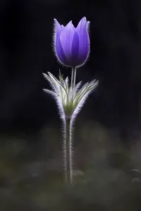
Trichome: Nature's Tiny Marvels Revealed. Step into the mesmerizing world of trichomes, where Botanik Digitalis purpurea L


All Professionally Made to Order for Quick Shipping
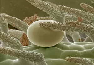
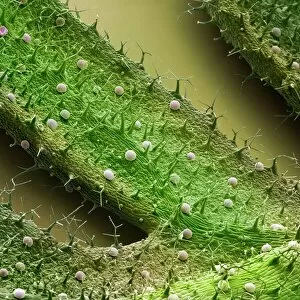
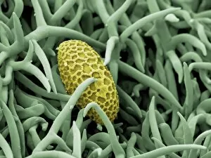
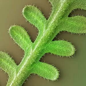
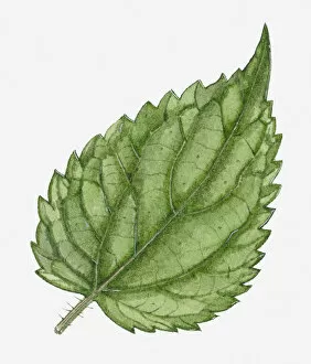
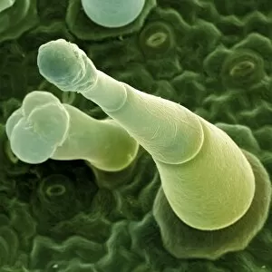
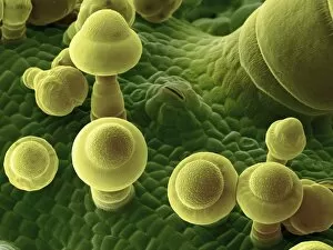
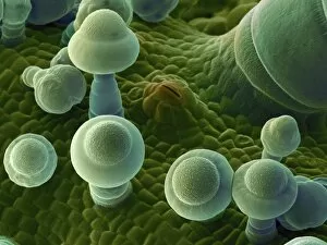
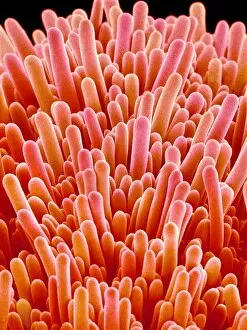
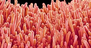
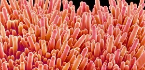
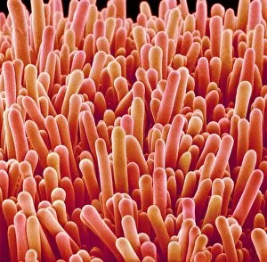
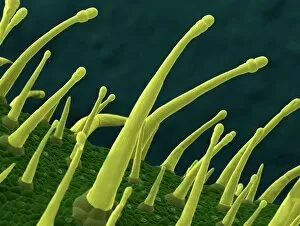
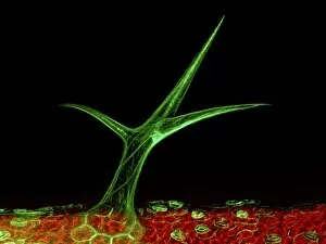
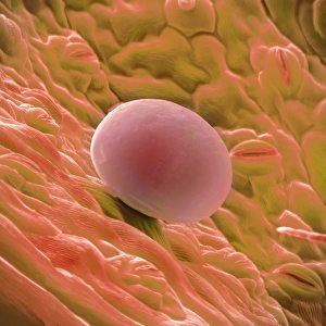
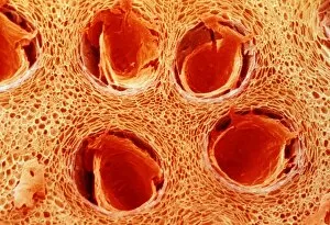
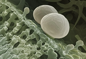
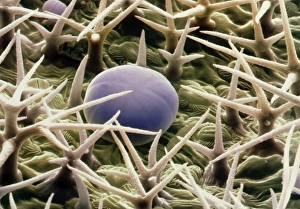
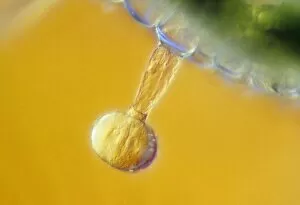
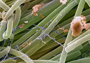
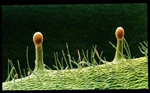
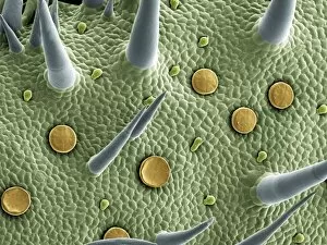
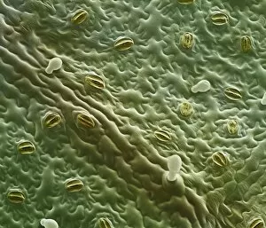
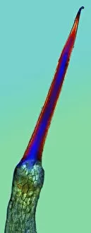
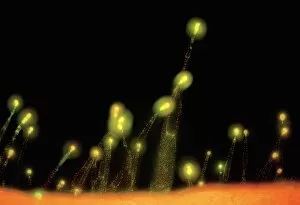
Trichome: Nature's Tiny Marvels Revealed. Step into the mesmerizing world of trichomes, where Botanik Digitalis purpurea L. Fingerhut 160:1 and other fascinating specimens await your exploration. These microscopic structures hold secrets that will leave you in awe. Take a closer look at the Thyme leaf oil gland, a tiny powerhouse responsible for its aromatic essence. Delicate and intricate, it is nature's gift to our senses. French lavender leaf surface captured under SEM reveals an enchanting landscape of trichomes. Like velvety hairs, they adorn the leaves, adding texture and beauty to this fragrant herb. Marvel at the Aubergine flower petal as seen through SEM lenses. Its surface showcases an array of trichomes standing tall like sentinels, protecting this delicate bloom from harm. Witness the captivating dance between Lily pollen grain and rosemary leaf under SEM magnification. A chance encounter showcasing nature's interconnectedness in stunning detail. Gorse flower bud unravels its hidden charm when observed through SEM lenses. Trichomes emerge like miniature spikes on its surface, providing protection while exuding elegance. Leaf oil glands come alive under SEM scrutiny – their unique shapes resembling precious jewels scattered across foliage, and are reservoirs of potent oils that contribute to plant health and vitality. French lavender leaf unveils its intricate tapestry when examined closely with SEM technology. Trichomes create a textured mosaic that adds depth to this beloved herb's allure. Periwinkle petal surface takes center stage as we delve into its microscopic realm using SEM imaging techniques. Trichomes delicately carpet this floral canvas, enhancing both aesthetics and functionality. Aubergine flower petals reveal their secret beauty through high-resolution SEM images – each trichome meticulously placed by nature to protect these vibrant blooms from external threats. Nettle leaf trichomes take on a life of their own when observed under SEM lenses.