Fraud Blocker Skip to main content

Transverse Collection (page 4)

"Exploring the Transverse World: From Subatomic Particles to Botanical Wonders" Unveiling the Secrets: The Higgs Boson Event captured by the ATLAS detector C013/6892

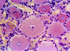
Background imageTransverse Collection: Ovarian follicles, light micrograph

Ovarian follicles, light micrograph
Ovarian follicles. Coloured light micrograph of a section through an ovary (orange), showing three primary follicles (pink)

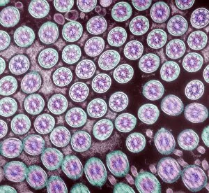
Background imageTransverse Collection: Cilia in cross section

Cilia in cross section
Cilia. Coloured transmission electron micrograph (TEM) of a cross section through cilia (circles), from the lining (epithelium) of the human trachea, or windpipe

Background imageTransverse Collection: Kidney glomeruli, light micrograph

Kidney glomeruli, light micrograph
Kidney glomeruli. Light microscope of a section through the cortex of a healthy kidney showing glomeruli. The core of a glomerulus is formed of a tightly coiled capillary (purple)

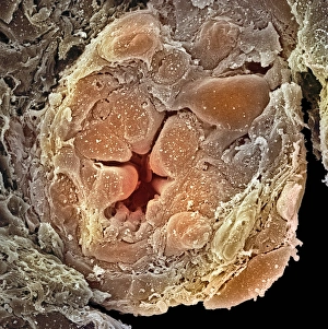
Background imageTransverse Collection: Collecting duct from a kidney, SEM

Collecting duct from a kidney, SEM
Collecting duct from a kidney. Coloured scanning electron micrograph (SEM) of a section through a collecting duct from a kidney

Background imageTransverse Collection: Salivary gland, light micrograph

Salivary gland, light micrograph
Salivary gland. Coloured light micrograph of a section through a sublingual salivary gland, which is situated directly under the tongue

Background imageTransverse Collection: Taste bud, light micrograph

Taste bud, light micrograph
Taste bud. Coloured light micrograph of a section through the tongue, showing a taste bud. This taste bud is seen as a round purple mass slightly to the right of the vertical white line

Background imageTransverse Collection: Spinal nerve ganglion, light micrograph

Spinal nerve ganglion, light micrograph
Spinal nerve ganglion. Light micrograph of a cross-section through a spinal nerve ganglion. This is a node of nerve cells located just outside the spinal cord at the point where it is joined by

Background imageTransverse Collection: Brain scan, artwork

Brain scan, artwork
Brain scan. Computer artwork based on an axial (cross-sectional) MRI scan of a human head, showing the brain. At upper left is the cerebellum (turquoise)

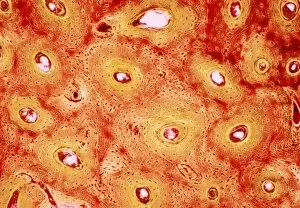
Background imageTransverse Collection: Bone canals, light micrograph

Bone canals, light micrograph
Bone canals. Coloured light micrograph of a section through human compact bone, showing Haversian canals (circular regions). The concentric rings surrounding the Haversian canals are called lamellae

Background imageTransverse Collection: Alcoholic hepatitis, light micrograph

Alcoholic hepatitis, light micrograph
Alcoholic hepatitis. Coloured light micrograph of a section through the liver of a patient with alcoholic hepatitis, inflammation of the liver due to prolonged heavy alcohol consumption

Background imageTransverse Collection: Normal brain, CT scans

Normal brain, CT scans
Normal brain. Coloured axial (cross-sectional) computed tomography (CT) scans of a healthy human head. The front of the head is at right in each scan

Background imageTransverse Collection: Normal brain, CT scan

Normal brain, CT scan
Normal brain, coloured axial (cross-sectional) computed tomography (CT) scan. The front of the head is at top. The brain is yellow (lower centre)

Background imageTransverse Collection: Cardiac muscle, SEM

Cardiac muscle, SEM
Cardiac muscle. Coloured scanning electron micrograph (SEM) of a bundle of cardiac muscle fibrils (green) from a healthy heart. Mitochondria (round, orange) supply the muscle cells with energy

Background imageTransverse Collection: Cerebellum structure, light micrograph

Cerebellum structure, light micrograph
Cerebellum structure. Coloured light micrograph of a section through the highly-folded cerebellum of the brain. The cerebellum comprises three main layers
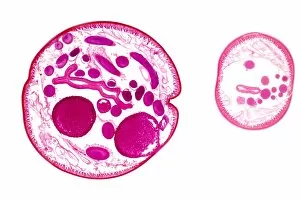
Background imageTransverse Collection: Ascaris roundworm, light micrograph

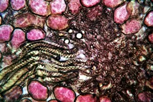
Ascaris roundworm, light micrograph

Background imageTransverse Collection: Grape vine stem, light micrograph

Grape vine stem, light micrograph
Grape vine stem. Polarised light micrograph of a cross-section through a young stem from the grape vine (Vitis vinifera). Most of the stem is a large central pith made up of parenchyma tissue (blue)

Background imageTransverse Collection: Water parsnip rhizome, light micrograph

Water parsnip rhizome, light micrograph
Water parsnip rhizome. Light micrograph of a cross-section through an underwater rhizome of the lesser water parsnip (Berula erecta)

Background imageTransverse Collection: Spinal cord, light micrograph

Spinal cord, light micrograph
Spinal cord. Light micrograph of a cross-section through a mammalian spinal cord. The central canal (white) is surrounded by dorsal (light brown) and ventral horns (dark brown)

Background imageTransverse Collection: Ovarian follicle, light micrograph

Ovarian follicle, light micrograph
Ovarian follicle. Light micrograph of a transverse section through a mammalian ovary, the reproductive structure that produces the female sex cells (ova)

Background imageTransverse Collection: Tree growth rings, light micrograph

Tree growth rings, light micrograph
Tree growth rings. Polarised light micrograph of a cross-section through a woody stem, showing a portion of an annual ring of xylem tissue with periods of seasonal growth

Background imageTransverse Collection: Mammalian ovary, light micrograph

Mammalian ovary, light micrograph
Mammalian ovary. Light micrograph of a transverse section through a mammalian ovary, the reproductive structure that produces the female sex cells (ova)

Background imageTransverse Collection: Virginia creeper stem, light micrograph

Virginia creeper stem, light micrograph
Virginia creeper stem. Polarised light micrograph of a cross-section through a stem from a Virginia creeper (Parthenocissus tricuspidatus)

Background imageTransverse Collection: Pine tree needle, light micrograph

Pine tree needle, light micrograph
Pine tree needle. Polarised light micrograph of a cross-section through a needle from a Pinus pine tree. This leaf is needle-like in order to reduce water loss (transpiration)

Background imageTransverse Collection: Celery stalk, light micrograph

Celery stalk, light micrograph
Celery stalk. Light micrograph of a cross-section through a stalk from a celery plant (Apium graveolens). Red eosin dye has been used to stain the xylem tissues

Background imageTransverse Collection: Tree-of-heaven stem, light micrograph

Tree-of-heaven stem, light micrograph
Tree-of-heaven stem. Polarised light micrograph of a cross-section through the stem of the tree-of-heaven (Ailanthus glandulosa). Below the outer layers (red) is a ring of vascular bundles

Background imageTransverse Collection: Aralia leaf stalk, light micrograph

Aralia leaf stalk, light micrograph
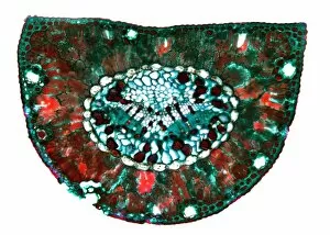
Aralia leaf stalk. Light micrograph of a cross-section through a leaf stalk (petiole) from an Araliatree. The outer layer directly below the epidermis is a supporting collenchyma ring (red)

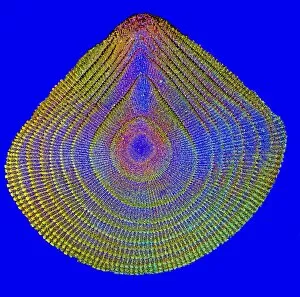
Background imageTransverse Collection: Sea urchin spine, light micrograph

Sea urchin spine, light micrograph
Sea urchin spine. Polarised light micrograph of a cross-section through a spine from an Echinus sea urchin. Sea urchin spines consist of supporting calcareous radial plates

Background imageTransverse Collection: Bracken fern rhizome, light micrograph

Bracken fern rhizome, light micrograph
Bracken fern rhizome. Polarised light micrograph of a cross-section through a rhizome from a bracken fern (Pteris aquilinum). The outer layer (yellow-green) consists of supporting fibres

Background imageTransverse Collection: Anatomical planes of the body, artwork

Anatomical planes of the body, artwork. The three planes shown here are the axial, saggital and coronal. The axial, or transverse, plane splits the body along a horizontal axis

Background imageTransverse Collection: Vascular disease progression, artwork

Vascular disease progression, artwork
Vascular disease progression. Artwork showing the progression of vascular disease in cross-section (left) and transverse section (right). At top is a healthy artery

Background imageTransverse Collection: Vascular anatomy, historical artwork

Vascular anatomy, historical artwork. 19th Century hand coloured lithographic print showing the arteries (red) and muscle structure (brown) of the human neck and chest

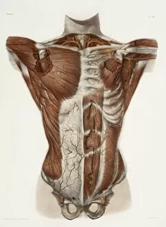
Background imageTransverse Collection: Human arteries, 19th Century illustration

Human arteries, 19th Century illustration. Historical hand coloured lithographic print showing the arteries (red) and muscle structure (brown) of the human torso

Background imageTransverse Collection: Burdock stem, light micrograph

Burdock stem, light micrograph
Burdock stem. Polarised light micrograph of a cross-section through the stem of a burdock plant (Arctium lappa). The outer layers below the epidermis are the collenchyma (red patches)

Background imageTransverse Collection: Brain structures, artwork

Brain structures, artwork
Brain structures. Computer artwork of a transverse section through a human brain showing a number of its regions. See image: C007/0802 for the labelled diagram

Background imageTransverse Collection: Moss stems, light micrograph

Moss stems, light micrograph. Transverse section through stems of a cord moss (Polytrichum commune). The outside of the stems, the epidermis, is made up of thick-walled cells

Background imageTransverse Collection: Fern frond, light micrograph

Fern frond, light micrograph. Transverse section through the frond of a fern (Cyrtomium falcatum), showing the sporangia. At left is a central vascular bundle (meristele)

Background imageTransverse Collection: Fern stem, light micrograph

Fern stem, light micrograph. Transverse section through a rachis (stem) of the bracken fern (Pteridium aquilinum). Under the outer epidermis (black) is a thin cortex (deep red)

Background imageTransverse Collection: Liverwort spore elaters, light micrograph

Liverwort spore elaters, light micrograph. Transverse section through the sporangium of a liverwort (Pellia epiphylla). This is the basal portion of the sporangium


All Professionally Made to Order for Quick Shipping

"Exploring the Transverse World: From Subatomic Particles to Botanical Wonders" Unveiling the Secrets: The Higgs Boson Event captured by the ATLAS detector C013/6892. Frozen in Time: Muybridge's iconic study of Horses Galloping reveals their transverse motion. Nature's Engineering Marvels: Witnessing the intricate Vascular Bundle through a SEM image. Beneath the Surface: A closer look at Compact Bone structure under a light micrograph. Revving Up History: Mechanics examine Bugatti Type 251 engine during the 1956 French Grand Prix. Rooted in Life: Maize Root depicted in stunning detail under a light micrograph. Battling Disease: Light micrograph showcases Liver Tissue Cirrhosis, revealing its transverse impact on health. Pancreatic Powerhouse: Islet of Langerhans illuminated through a captivating light micrograph. Hidden Depths Below Ground: Exploring Water Fern Rhizome's intricate network using a light micrograph technique. Tall and Strong Guardians of Nature - Pine Tree Stem revealed through an enchanting light micrograph image Xylem Tissue Exposed. Delicate structures captured with precision using SEM technology Wood Underneath Our Feet.