
Trabecular Collection
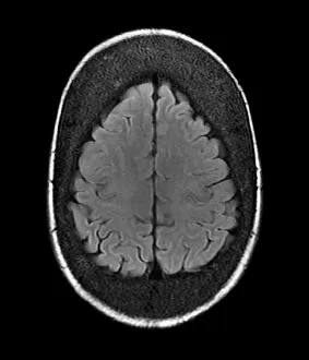
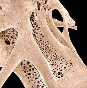
"Trabecular: Unveiling the Intricate World of Bone Structure" Have you ever wondered what lies beneath our thickened skull


All Professionally Made to Order for Quick Shipping
"Trabecular: Unveiling the Intricate World of Bone Structure" Have you ever wondered what lies beneath our thickened skull? Thanks to advanced medical technology like MRI scans, we can now explore the fascinating world bone. These intricate patterns within our skull not only provide strength and support but also play a crucial role in protecting our delicate brain. Delving deeper into the realm of bone structure, artists have beautifully depicted the complexity formations through their artwork. Their creations offer us a glimpse into this hidden network that forms the foundation of our skeletal system. Examining bone tissue under a scanning electron microscope (SEM) reveals an astonishing level of detail. The magnified images showcase the interwoven nature of trabeculae, resembling an elaborate web that ensures optimal strength while minimizing weight. Not limited to humans alone, even fish bones exhibit similar intricacies when observed under SEM. Each tiny fragment showcases its unique arrangement, highlighting nature's remarkable design principles. Moving beyond skulls and fish bones, X-ray imaging allows us to appreciate other parts of our body where trabecular structures are present. A thigh bone head captured on an X-ray reveals how these bony networks extend throughout different regions, providing stability and flexibility for movement. These captivating glimpses into trabecular bone remind us that there is so much more than meets the eye when it comes to understanding our own anatomy. From thickened skulls revealed by MRI scans to detailed SEM images capturing bone tissue at microscopic levels – each image tells a story about the incredible architecture that supports us every day.