
Thrombosis Collection
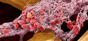
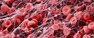
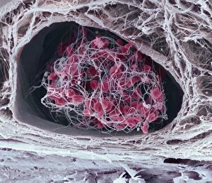
"Thrombosis: The Silent Threat Within" Blood clot, SEM C016 / 9747 - a microscopic view of the intricate web that can lead to danger within our veins


All Professionally Made to Order for Quick Shipping
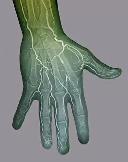
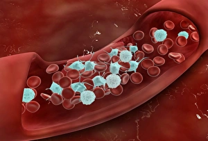
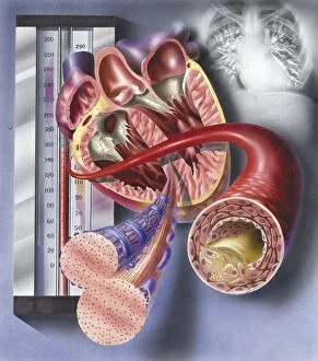
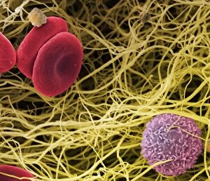
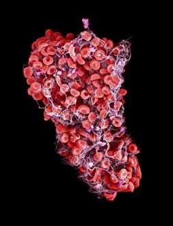
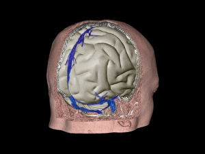
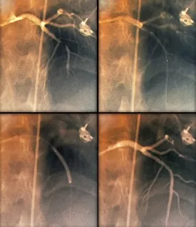
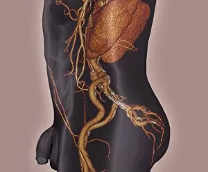
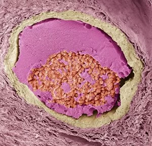
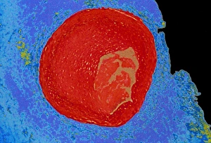
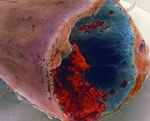
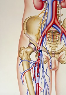
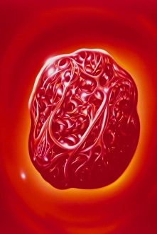
"Thrombosis: The Silent Threat Within" Blood clot, SEM C016 / 9747 - a microscopic view of the intricate web that can lead to danger within our veins. Heart shape in clouds, conceptual image - a reminder that even something as beautiful as love can be affected by the lurking threat of thrombosis. Blood clot, SEM - an up-close look at the culprit behind this potentially life-threatening condition. Gangrene from arterial thrombosis following embolism (colour litho) - a haunting visual representation of the consequences when blood flow is blocked and tissues are deprived of oxygen. Ischaemia, digital angiogram - revealing how they are disrupt blood supply to vital organs, leading to tissue damage and potential organ failure. Blood clot, artwork C016 / 4619 - an artistic interpretation capturing both the beauty and danger intertwined within our circulatory system. Thrombosed blood vessel, artwork C013 / 4649 - showcasing the dangerous aftermath when a blood vessel becomes obstructed by a menacing clot. Arteries on heart showing atherosclerotic plaque in an artery - highlighting how buildup within our arteries can contribute to increased risk for thrombotic events. Microscopic view of blood clotting inside the artery - unveiling the complex process where platelets aggregate and fibrin forms clots that may have devastating consequences if left untreated. Acute coronary syndrome – microvascular obstruction – illustrating how even small blockages caused by clots can result in significant damage to cardiac tissue and potentially trigger heart attacks or strokes. Interior view of heart with detail of muscle cells and atherosclerotic artery – emphasizing how thrombosis not only affects circulation but also impacts vital cardiac structures essential for proper functioning. Thrombus forming on valve within vein – shedding light on one possible location where these dangerous clots may form silently until complications arise.