
Thrombosed Collection
"Unveiling the Intricacies of Thrombosed: From Blood Vessels to Brain and Beyond" Discover the captivating world conditions through a series of remarkable medical images


All Professionally Made to Order for Quick Shipping
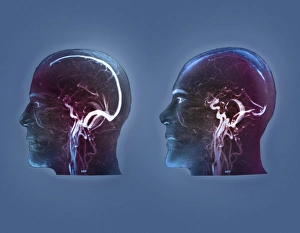
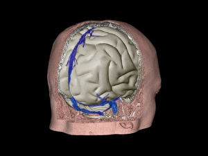
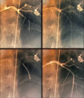
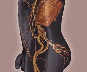
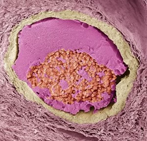
"Unveiling the Intricacies of Thrombosed: From Blood Vessels to Brain and Beyond" Discover the captivating world conditions through a series of remarkable medical images. Artwork C013 / 4649 showcases the intricate nature of a thrombosed blood vessel, highlighting its complexity and impact on our health. Delving deeper into this realm, MRI scans reveal the presence of thrombophlebitis in the brain, shedding light on the potential consequences and emphasizing the need for timely intervention. Witnessing coronary stenosis before treatment via X-ray imagery reminds us of the challenges faced by individuals with this condition. However, hope emerges as we witness coronary stenosis after treatment in subsequent X-rays, showcasing medical advancements that restore normalcy to affected hearts. The journey continues as 3D CT scans unveil another dimension of understanding thrombophlebitis in the brain. These detailed images provide invaluable insights into diagnosis and guide effective treatments. Returning to coronary stenosis once more, we witness both pre- and post-treatment scenarios through X-ray examinations. The transformative power of medical interventions becomes evident as these images showcase improved heart health following successful procedures. Concluding our exploration is an intriguing glimpse into atherosclerosis captured by a 3D CT scan. This image serves as a reminder that vigilance against thrombotic conditions extends beyond specific areas; it encompasses our entire cardiovascular system. These mesmerizing visuals not only educate but also inspire awe at human ingenuity in unraveling complex medical mysteries. They remind us that behind every diagnosis lies an opportunity for progress – be it through innovative treatments or enhanced preventive measures. Let us continue to explore, understand, and combat thrombosed conditions together for healthier lives ahead.