Fraud Blocker Skip to main content

Thrombocytes Collection

Thrombocytes, also known as platelets, play a crucial role in our body's defense against bleeding

Background imageThrombocytes Collection: Blood clot, SEM C016 / 9747

Blood clot, SEM C016 / 9747
Blood clot, coloured scanning electron micrograph (SEM). Red blood cells (erythrocytes) are trapped within a fibrin protein mesh (beige)

Background imageThrombocytes Collection: White blood cells and platelets, SEM C016 / 3099

White blood cells and platelets, SEM C016 / 3099
White blood cells and platelets. Coloured scanning electron micrograph (SEM) of platelets (green) with a variety of different white blood cells (leucocytes, yellow)

Background imageThrombocytes Collection: White blood cells and platelets, SEM C016 / 3098

White blood cells and platelets, SEM C016 / 3098
White blood cells and platelets. Coloured scanning electron micrograph (SEM) of platelets (purple) with a variety of different white blood cells (leucocytes, blue)

Background imageThrombocytes Collection: Blood clot, SEM C016 / 9751

Blood clot, SEM C016 / 9751
Blood clot, coloured scanning electron micrograph (SEM). Red blood cells (erythrocytes) are trapped within a fibrin protein mesh (beige)

Background imageThrombocytes Collection: Blood clot, SEM C016 / 9746

Blood clot, SEM C016 / 9746
Blood clot, coloured scanning electron micrograph (SEM). Red blood cells (erythrocytes) are trapped within a fibrin protein mesh (beige)

Background imageThrombocytes Collection: Blood clot, SEM C016 / 9749

Blood clot, SEM C016 / 9749
Blood clot, coloured scanning electron micrograph (SEM). Red blood cells (erythrocytes) are trapped within a fibrin protein mesh (beige)

Background imageThrombocytes Collection: Blood clot, SEM C016 / 9750

Blood clot, SEM C016 / 9750
Blood clot, coloured scanning electron micrograph (SEM). Red blood cells (erythrocytes) are trapped within a fibrin protein mesh (beige)

Background imageThrombocytes Collection: Blood clot, SEM C016 / 9753

Blood clot, SEM C016 / 9753
Blood clot. Coloured scanning electron micrograph (SEM) of a blood clot from the inner wall of the left ventricle of a heart

Background imageThrombocytes Collection: Blood clot, SEM C016 / 9752

Blood clot, SEM C016 / 9752
Blood clot, coloured scanning electron micrograph (SEM). Red blood cells (erythrocytes) are trapped within a fibrin protein mesh (beige)

Background imageThrombocytes Collection: Blood clot, SEM C016 / 9745

Blood clot, SEM C016 / 9745
Blood clot, coloured scanning electron micrograph (SEM). Red blood cells (erythrocytes) are trapped within a fibrin protein mesh (beige)

Background imageThrombocytes Collection: Blood clot, SEM C016 / 9748

Blood clot, SEM C016 / 9748
Blood clot, coloured scanning electron micrograph (SEM). Red blood cells (erythrocytes) are trapped within a fibrin protein mesh (beige)

Background imageThrombocytes Collection: Blood clot, SEM C015 / 9608

Blood clot, SEM C015 / 9608
Blood clot. Coloured scanning electron micrograph (SEM) of a blood clot from the inner wall of the left ventricle of a heart

Background imageThrombocytes Collection: Blood clot, SEM C016 / 9744

Blood clot, SEM C016 / 9744
Blood clot, coloured scanning electron micrograph (SEM). Red blood cells (erythrocytes) are trapped within a fibrin protein mesh (beige)

Background imageThrombocytes Collection: Blood clot, SEM C014 / 0381

Blood clot, SEM C014 / 0381
Blood clot. Coloured scanning electron micrograph (SEM) of a blood clot (thrombus) in an arteriole (small blood vessel) of a salivary gland

Background imageThrombocytes Collection: Blood clot, SEM C014 / 0380

Blood clot, SEM C014 / 0380
Blood clot. Coloured scanning electron micrograph (SEM) of a blood clot (thrombus) in an arteriole (small blood vessel) of a salivary gland

Background imageThrombocytes Collection: White blood cells, SEM C016 / 3102

White blood cells, SEM C016 / 3102
White blood cells. Coloured scanning electron micrograph (SEM) of white blood cells (leucocytes) from a sample of peripheral blood

Background imageThrombocytes Collection: Blood cells, SEM C016 / 3101

Blood cells, SEM C016 / 3101
Blood cells. Coloured scanning electron micrograph (SEM) of a variety of different white blood cells (leucocytes, blue/purple) with two red blood cells (erythrocytes)

Background imageThrombocytes Collection: Blood cells, SEM C016 / 3100

Blood cells, SEM C016 / 3100
Blood cells. Coloured scanning electron micrograph (SEM) of a variety of different white blood cells (leucocytes, yellow/green) with two red blood cells (erythrocytes)

Background imageThrombocytes Collection: Macrophage and platelets, SEM C016 / 3094

Macrophage and platelets, SEM C016 / 3094
Macrophage and platelets. Coloured scanning electron micrograph (SEM) of platelets (green) attached to a macrophage white blood cell (turquoise)

Background imageThrombocytes Collection: Platelets, SEM C016 / 3091

Platelets, SEM C016 / 3091
Platelets, coloured scanning electron micrograph (SEM). Platelets, or thrombocytes, are fragments of white blood cells. When blood clots

Background imageThrombocytes Collection: Blood clot C013 / 7129

Blood clot C013 / 7129
Blood clot. Coloured scanning electron micrograph (SEM) of a blood clot (centre) forming in connective tissue. Red blood cells (erythrocytes)

Background imageThrombocytes Collection: Blood clot C013 / 7128

Blood clot C013 / 7128
Blood clot. Coloured scanning electron micrograph (SEM) of a blood clot (centre) forming in connective tissue. Red blood cells (erythrocytes)

Background imageThrombocytes Collection: Blood cells, artwork C013 / 4726

Blood cells, artwork C013 / 4726
Blood cells. Computer artwork of red blood cells (erythrocytes, red), white blood cells (leucocytes, white), and platelets (thrombocytes, yellow)

Background imageThrombocytes Collection: Blood cells, artwork C013 / 4707

Blood cells, artwork C013 / 4707
Blood cells. Computer artwork of red blood cells (erythrocytes, red), white blood cells (leucocytes, white), and platelets (thrombocytes, yellow)

Background imageThrombocytes Collection: Platelets in a blood clot

Platelets in a blood clot. Coloured scanning electron micrograph (SEM) of platelet cells (thrombocytes) in a blood clot. They are oval shaped

Background imageThrombocytes Collection: Blood platelet, TEM

Blood platelet, TEM

Background imageThrombocytes Collection: Platelets, SEM

Platelets, SEM
Platelets. Coloured scanning electron micrograph (SEM) of a clump of activated platelets from a blood clot. Platelets are fragments of white blood cells

Background imageThrombocytes Collection: Activated platelets, SEM

Activated platelets, SEM
Activated platelets. Coloured scanning electron micrograph (SEM) of activated platelets, or thrombocytes, starting to adhere to exposed sub-endothelium on the inner surface of a vein

Background imageThrombocytes Collection: Platelet activation, artwork

Platelet activation, artwork
actication of PRP gel

Background imageThrombocytes Collection: Red blood cells and platelets, SEM

Red blood cells and platelets, SEM
Red blood cells and platelets. Coloured scanning electon micrograph (SEM) of human erythrocytes (red blood cells) and a platelet aggregate (purple)

Background imageThrombocytes Collection: Blood cells in blood vessel, artwork

Blood cells in blood vessel, artwork
Blood cells in blood vessel, computer artwork. Red blood cells (erythrocytes, red) distribute oxygen to body tissues, and carry waste carbon dioxide back to the lungs

Background imageThrombocytes Collection: Human blood cells, SEM

Human blood cells, SEM
Human blood cells, coloured scanning electron micrograph (SEM). Seen here are platelets (small, roundish) and a neutrophil white blood cell (large, whitish)


All Professionally Made to Order for Quick Shipping

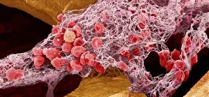
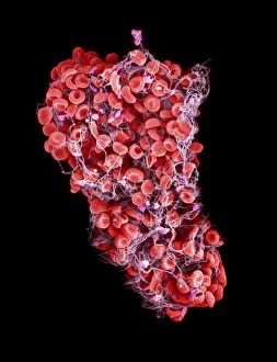
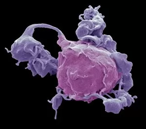
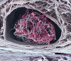
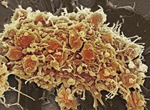
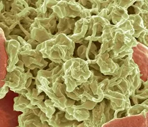
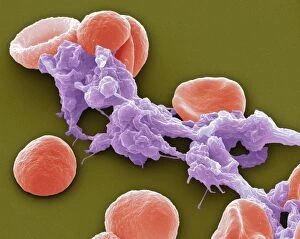
Thrombocytes, also known as platelets, play a crucial role in our body's defense against bleeding. These tiny cells are responsible for forming blood clots, which help to seal wounds and prevent excessive blood loss. In these stunning scanning electron microscope (SEM) images, we can observe the intricate structure up close. The first image (SEM C016 / 9747) showcases a single platelet surrounded by other blood components. Its irregular shape and granular appearance highlight its unique characteristics. Platelets are derived from megakaryocytes in the bone marrow and circulate in our bloodstream until they are activated upon injury or damage. Moving on to the second image (Platelets, SEM), we witness a cluster of platelets working together to form a clot. This remarkable process involves platelet adhesion and aggregation at the site of injury, creating a mesh-like network that traps red blood cells and forms a solid plug. In SEM C016 / 3099 and SEM C016 / 3098, we observe white blood cells alongside platelets. White blood cells play an essential role in immune response but can also contribute to clot formation when necessary. The collaboration between these two cell types ensures efficient wound healing while maintaining overall health. The remaining images depict various stages of blood clot formation: SEM C016 / 9751, SEM C016 / 9746, SEM C016 / 9749, SEM C016 / 9750, SEM C016 / 9753, SEM C016 / 9752 showcase different angles and magnifications capturing the intricate details of this vital process. From their initial activation to fibrin deposition resulting in stable clots (as shown in Blood Clot -SEM C016/9745 & Blood Clot -SEM-CO16/9748), thrombocytes demonstrate their indispensable role within our circulatory system.