
Taste Bud Collection
The taste bud: Unlocking the Secrets of Flavor Have you ever wondered how your tongue can detect different tastes


All Professionally Made to Order for Quick Shipping
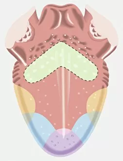
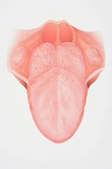
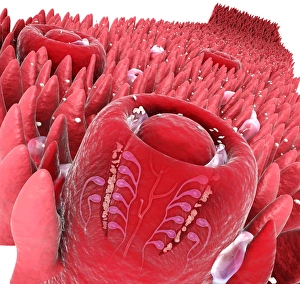
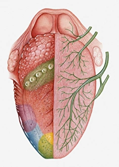
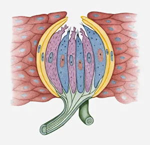
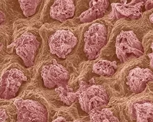
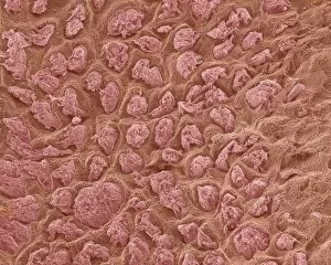
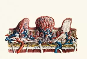
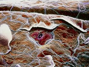
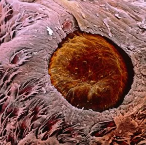
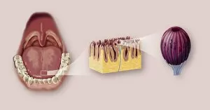
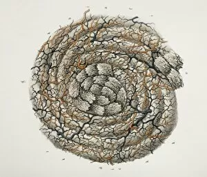
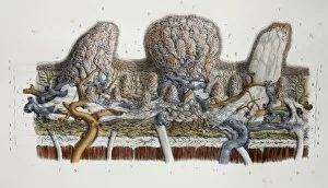
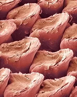
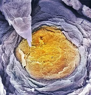
The taste bud: Unlocking the Secrets of Flavor Have you ever wondered how your tongue can detect different tastes? The answer lies within the intricate world of taste buds. A taste map of a human tongue reveals fascinating insights into our sense of taste. In this map, we see distinct areas highlighted in various colors. The light green area is particularly sensitive to bitter tastes, while yellow areas are attuned to sour flavors. Blue regions on the tongue respond to saltiness, and a purple area is dedicated to detecting sweetness. Examining the anatomy of the tongue from a front view provides further clarity. A digital cross-section illustration showcases taste buds alongside filiform papillae, which play a crucial role in texture perception. Delving deeper into these microscopic structures, an illustration displays a cross-section of a human taste bud itself. This visual representation allows us to appreciate its complexity and understand how it contributes to our sensory experiences. Moving beyond individual taste buds, an illustration highlights various sections on the surface of the human tongue that correspond with specific tastes. These pink-highlighted areas indicate where our gustatory receptors reside alongside fungiform papillae – tiny bumps responsible for housing many tastebuds. To comprehend how these sensations travel through our body, another cross-sectional image depicts both cranial nerve pathways and designated regions for tasting on one side of the human tongue's cross-section. While humans aren't alone in possessing this remarkable ability, dogs also have their own unique tongues with similar anatomical features as depicted in an illustrated cross-section specifically showcasing canine tongues. Biomedical illustrations provide additional insight by pinpointing not only where exactly tastebuds are located but also revealing their intricate structure at close range. Ultimately, understanding these complex mechanisms enhances our appreciation for flavor perception. By examining detailed images depicting both external and internal aspects related to tasting – including connections with smell and touch – we gain valuable knowledge about one of life's most delightful senses: Taste.