Fraud Blocker Skip to main content

Swelling Collection

"Swelling: A Visual Journey into the World of Medical Conditions" Bunions and X-ray

Background imageSwelling Collection: Bunions, X-ray

Bunions, X-ray
Bunions. Coloured X-ray of an elderly womans deformed inward-turning toes due to bunions (orange, lower centre). This deformity (hallux valgus) is caused by wearing ill-fitting shoes

Background imageSwelling Collection: Tendinitis of the shoulder, X-ray

Tendinitis of the shoulder, X-ray
Tendinitis of the shoulder. Coloured X-ray of the shoulder of a patient showing calcification (grainy patches) around the rotator cuff tendon (upper left)

Background imageSwelling Collection: Bacterial meningitis, MRI scan

Bacterial meningitis, MRI scan
Bacterial meningitis. Coloured magnetic resonance imaging (MRI) scan of an axial section through the brain of a 16-year-old patient

Background imageSwelling Collection: Slipped disc

Slipped disc. Spinal model demonstrating a slipped intervertebral disc. The slipped disc is the small bulge (red, centre), near the base of the spine

Background imageSwelling Collection: RF- Pool Frog (Rana lessonae) sitting on white lily pad, Danube delta rewilding area, Romania

RF- Pool Frog (Rana lessonae) sitting on white lily pad, Danube delta rewilding area, Romania

Background imageSwelling Collection: Comic birthday postcard, Boy with large cake, and a swelling stomach Date

Comic birthday postcard, Boy with large cake, and a swelling stomach Date: 20th century

Background imageSwelling Collection: Fungus of the dura mater, from Selecta Praxis Medico-Chirurgica

Fungus of the dura mater, from Selecta Praxis Medico-Chirurgica
CHT234598 Fungus of the dura mater, from Selecta Praxis Medico-Chirurgica by Alexander Auvert, Paris, 1856 (coloured engraving) by French School

Background imageSwelling Collection: Diseases of the Stomach and Kidneys (colour litho)

Diseases of the Stomach and Kidneys (colour litho)
6006706 Diseases of the Stomach and Kidneys (colour litho) by European School, (19th century); Private Collection; (add.info.: Diseases of the Stomach and Kidneys)

Background imageSwelling Collection: Gout (engraving)

Gout (engraving)
3646416 Gout (engraving) by Burgkmair, Hans (c.1473-1531) (after); Private Collection; (add.info.: Gout. Woodcut from a series depicting physical infirmities)

Background imageSwelling Collection: Cartoon commenting on the land acquisitions of the United States of America

Cartoon commenting on the land acquisitions of the United States of America
CHT206524 Cartoon commenting on the land acquisitions of the United States of America, illustration from Le Rire, 4th March 1895 (litho) (b/w photo) by French School

Background imageSwelling Collection: Achilles contending with the Rivers (engraving)

Achilles contending with the Rivers (engraving)
973689 Achilles contending with the Rivers (engraving) by Flaxman, John (1755-1826) (after); Private Collection; (add.info.: Achilles contending with the Rivers)

Background imageSwelling Collection: 'Look, Look, Near the Vistula. The German Bellies are Swelling Up

"Look, Look, Near the Vistula. The German Bellies are Swelling Up
1068611 " Look, Look, Near the Vistula. The German Bellies are Swelling Up. So They Don t Feel So Good", 1914-15 (colour litho) by Malevich

Background imageSwelling Collection: Sunbathing on a balcony to treat chronic inflammation of the knee (engraving)

Sunbathing on a balcony to treat chronic inflammation of the knee (engraving)
3097144 Sunbathing on a balcony to treat chronic inflammation of the knee (engraving) by German School, (19th century); Private Collection; (add.info)

Background imageSwelling Collection: Thyroid disease, from a book by Baron Jean Louis Alibert (1768-1837

Thyroid disease, from a book by Baron Jean Louis Alibert (1768-1837
CHT235328 Thyroid disease, from a book by Baron Jean Louis Alibert (1768-1837) 1817 (coloured engraving) by French School, (19th century); Bibliotheque de la Faculte de Medecine, Paris

Background imageSwelling Collection: Cestina Warehouse or Belly Piece Shop, pub. 1793 (hand coloured engraving)

Cestina Warehouse or Belly Piece Shop, pub. 1793 (hand coloured engraving)
5902917 Cestina Warehouse or Belly Piece Shop, pub. 1793 (hand coloured engraving) by Cruikshank, Isaac (1756-1811); Private Collection; (add.info)

Background imageSwelling Collection: Twilight at Varese (chromolitho)

Twilight at Varese (chromolitho)
5222048 Twilight at Varese (chromolitho) by von Cramm, Helga (1840-1919); Private Collection; (add.info.: Twilight at Varese)

Background imageSwelling Collection: Guam: A Man with Elephantiasis, from Voyage Autour du Monde sur les Corvettes de

Guam: A Man with Elephantiasis, from Voyage Autour du Monde sur les Corvettes de
TRV173970 Guam: A Man with Elephantiasis, from Voyage Autour du Monde sur les Corvettes de L Uranie 1817-20 engraved by Choubard, published 1825 (engraving) by Arago

Background imageSwelling Collection: Joe Frazier seen here giving a press conference whilst an aide applies a ice bag to his

Joe Frazier seen here giving a press conference whilst an aide applies a ice bag to his face to reduce the swelling following the fight against Muhammad Ali

Background imageSwelling Collection: Henry Cooper, the new British and Empire Heavyweight boxing champion looks from behind

Henry Cooper, the new British and Empire Heavyweight boxing champion looks from behind dark glasses at the annual Variety Club of Great Britain sports celebrities lunch at the Savoy

Background imageSwelling Collection: Marie Carteri, 1737, (1885)

Marie Carteri, 1737, (1885). Cured on 4 September at the tomb of M[?] de Paris, all pain ceased. The next morning, on removing her bandage or blindfold

Background imageSwelling Collection: A man on a white horse, whom Cuthbert believed to be an angel

A man on a white horse, whom Cuthbert believed to be an angel, gave the saint advice on how to cure a swelling on his knee which had made walking impossible. Saint Cuthbert, c. 634 - 687

Background imageSwelling Collection: Waves swelling and breaking along the rocks and wall on the coast

Waves swelling and breaking along the rocks and wall on the coast; Hastings, England

Background imageSwelling Collection: Bulbous light hovering over Hessdalen, Norway

Bulbous light hovering over Hessdalen, Norway
This bulbous light is seen to swell in size while retaining something of a classic domed disc saucer configuration. Mysterious lights appear regularly over Hessdalen, Norway

Background imageSwelling Collection: Brain injury C016 / 8920

Brain injury C016 / 8920
Brain injury. Gross specimen of a section through a brain showing the effects of a blunt trauma. The hemisphere at right is showing extensive haemorrhaging (bleeding)

Background imageSwelling Collection: Heart with arteries showing cholesterol in one and plaque in the other

Heart with arteries showing cholesterol in one and plaque in the other
Heart with arteries showing cholesterol in one artery and atherosclerotic plaque in the other

Background imageSwelling Collection: Build-up of atherosclerotic plaque in an artery

Build-up of atherosclerotic plaque in an artery

Background imageSwelling Collection: Acute coronary syndrome - microvascular obstruction

Acute coronary syndrome - microvascular obstruction

Background imageSwelling Collection: Thrombus forming on valve within vein

Thrombus forming on valve within vein

Background imageSwelling Collection: Normal artery compared to plaque and thrombus formation in artery

Normal artery compared to plaque and thrombus formation in artery

Background imageSwelling Collection: Artery showing atherosclerotic plaque, platelets and red blood cells

Artery showing atherosclerotic plaque, platelets and red blood cells
Close-up of artery showing atherosclerotic plaque, platelets and red blood cells

Background imageSwelling Collection: Virus, swelling caused by Poliovirus, drawing

Virus, swelling caused by Poliovirus, drawing
Medicine - Anatomy - Microbiology - Infectious Diseases - Virology. Virus. Swelling caused by Poliovirus. Drawing

Background imageSwelling Collection: Drainage trocar with cannulae, circa 1860 C017 / 6949

Drainage trocar with cannulae, circa 1860 C017 / 6949
Dr. Southeys drainage trocar with 3 cannulae in their case. This set dates from 1860 and is made by Arnold and Sons, England

Background imageSwelling Collection: Drainage trocar with cannulae, circa 1860 C017 / 6950

Drainage trocar with cannulae, circa 1860 C017 / 6950
Dr. Southeys drainage trocar with 3 cannulae arranged on an orange background. This set dates from 1860 and is made by Arnold and Sons, England. The instruments are made from ivory and silver

Background imageSwelling Collection: Colitis, light micrograph

Colitis, light micrograph
Colitis. Light micrograph of a section through colon (large intestine) tissue affected by bacterial infection, causing injury to the mucosal lining, which has resulted in inflammation (colitis)

Background imageSwelling Collection: Nerve ganglion, light micrograph

Nerve ganglion, light micrograph
Nerve ganglion. Light micrograph of a section through a dorsal (sensory) spinal root ganglion associated with a sensory nerve root of the spinal cord. Sensory information from peripheral sites e.g

Background imageSwelling Collection: Oedema of larynx, artwork C016 / 6529

Oedema of larynx, artwork C016 / 6529
Oedema of larynx. Artwork of a transverse (axial) section through a larynx (seen from above) with and without swelling. The bottom image shows a healthy larynx

Background imageSwelling Collection: Oedema of larynx, artwork C016 / 6528

Oedema of larynx, artwork C016 / 6528
Oedema of larynx. Artwork of a vertical (coronal) section through a larynx, showing oedema (swelling caused by fluid accumulation)

Background imageSwelling Collection: Oedema of larynx, artwork C016 / 6530

Oedema of larynx, artwork C016 / 6530
Oedema of larynx. Artwork of sections through a larynx with and without swelling. The bottom image shows a healthy larynx in transverse (axial) section from above

Background imageSwelling Collection: Brain injury C016 / 8921

Brain injury C016 / 8921
Brain injury. Gross specimen of a section through a brain showing swelling and haemorrhaging (brown areas) in one hemisphere (right)

Background imageSwelling Collection: Arthritic hands, X-ray C018 / 0534

Arthritic hands, X-ray C018 / 0534
Arthritic hands. Coloured X-ray of the hands of a 72 year old patient with rheumatoid arthritis. The wrist and finger joints have become inflamed and swollen

Background imageSwelling Collection: Arthritic hands, X-ray C018 / 0532

Arthritic hands, X-ray C018 / 0532
Arthritic hands. X-ray of the hands of a 72 year old patient with rheumatoid arthritis. The wrist and finger joints have become inflamed and swollen

Background imageSwelling Collection: Trocar and cannula, circa 1860 C017 / 6960

Trocar and cannula, circa 1860 C017 / 6960
Trocar and cannula dating from 1860, England. The instruments are made from ivory, steel and silver. They were generally used to drain fluid-filled swellings

Background imageSwelling Collection: High endothelial venule, TEM C014 / 1446

High endothelial venule, TEM C014 / 1446
High endothelial venule. Transmission electron micrograph (TEM) of a section through a high endothelial venule (HEV). Venules are tiny veins that transport deoxygenated blood from the capillary beds

Background imageSwelling Collection: High endothelial venule, TEM C014 / 1445

High endothelial venule, TEM C014 / 1445
High endothelial venule. Transmission electron micrograph (TEM) of a section through a high endothelial venule (HEV). Venules are tiny veins that transport deoxygenated blood from the capillary beds

Background imageSwelling Collection: Plegmon eye inflammation, artwork

Plegmon eye inflammation, artwork
Plegmon eye inflammation. Artwork of a sagittal section through a human eye and the surrounding structures, in a case of a subcutaneous (below the skin) phlegmon inflammation of an eye cavity

Background imageSwelling Collection: Foreign body in finger, X-ray C017 / 8009

Foreign body in finger, X-ray C017 / 8009
Foreign body in finger. X-ray of the finger of a 60 year old male patient that contains a foreign body that is causing swelling of the soft tissue

Background imageSwelling Collection: Oedema of the hand C015 / 3430

Oedema of the hand C015 / 3430
Oedema of the hand. Close-up of swollen skin on the hand of a woman. Oedema is an abnormal swelling due to an accumulation of fluid in the body tissues

Background imageSwelling Collection: Physiotherapist treating gymnast C015 / 6203

Physiotherapist treating gymnast C015 / 6203
Sports medicine. A physiotherapist checking the ankles of a gymnast during a training session


All Professionally Made to Order for Quick Shipping

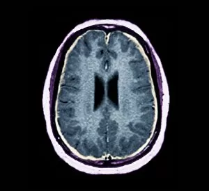
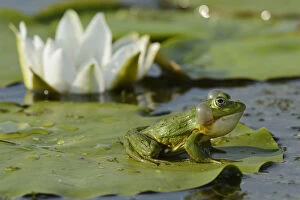
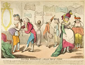
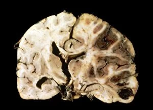
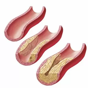
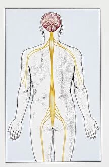
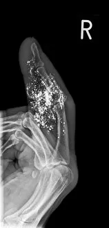
"Swelling: A Visual Journey into the World of Medical Conditions" Bunions and X-ray: Unveiling the Painful Swellings that Plague Feet Tendinitis of the Shoulder and X-ray: Understanding Inflammation in Motion Testicle Growth Drained: A Tale of Relief from an Uncomfortable Swelling Bacterial Meningitis and MRI Scan: Peering Inside the Swollen Brain's Battle Slipped Disc: When a Backache Turns into a Debilitating Swelling Elephantiasis and X-ray: Exploring Rare Cases of Extreme Swellings RF- Pool Frog on White Lily Pad, Danube Delta Rewilding Area, Romania - Nature's Serene Beauty Amidst Life's Many Swellings Comic Birthday Postcard with Boy, Cake, and a Swelling Stomach Date - Celebrating with Laughter (and Maybe Some Indigestion) Fungus of the Dura Mater from Selecta Praxis Medico-Chirurgica - Delving into Intriguing Cases of Cranial Swellings Diseases of the Stomach and Kidneys (Colour Litho) - Examining Internal Battles within our Vital Organs' Walls Gout (Engraving): The Agonizing Tale Behind Joint Inflammation Cartoon Commenting on Land Acquisitions in America.