
Striated Muscle Collection
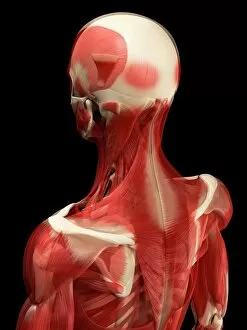
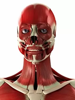
"Unleashing the Power Within: Exploring the Intricate World of Striated Muscle" In this captivating artwork, we delve into the awe-inspiring realm of male muscles


All Professionally Made to Order for Quick Shipping
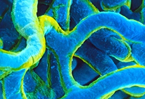
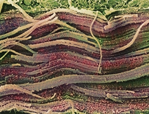
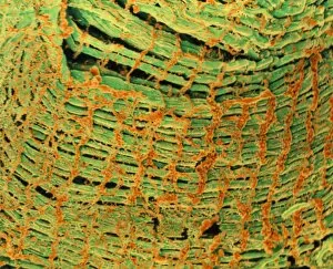
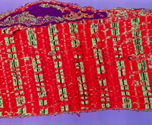
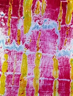
"Unleashing the Power Within: Exploring the Intricate World of Striated Muscle" In this captivating artwork, we delve into the awe-inspiring realm of male muscles. With intricate details and vibrant colors, it beautifully showcases the sheer strength and complexity that lies within our bodies. Through a false-color scanning electron microscope (SEM) image, we witness the remarkable connection between pericyte capillaries and striated muscle. This microscopic view unveils a hidden world where blood vessels intricately intertwine with muscular fibers, providing vital nourishment to these powerhouses. Zooming in further, we encounter an astonishing molecule called myosin fragment F006/9245. This molecular marvel plays a pivotal role in muscle contraction, enabling us to perform incredible feats of strength and endurance. The accompanying series of artwork takes us on a visual journey through male muscle structure. Each piece meticulously portrays various angles and perspectives, highlighting the sculpted contours and bulging sinews that define these powerful physiques. As we admire these artistic renderings, let's not forget the significance of striated muscles in our daily lives. From lifting heavy weights to running marathons or even performing simple tasks like walking or typing – every movement is made possible by their coordinated contractions, and are truly fascinating entities that deserve our admiration for their unwavering dedication to keeping us active and mobile. So let's celebrate this extraordinary gift bestowed upon us as we continue exploring the wonders hidden beneath our skin.