Fraud Blocker Skip to main content

Streptococcus Pneumoniae Collection

"Unveiling the Intricacies of Streptococcus pneumoniae

Background imageStreptococcus Pneumoniae Collection: Bacterial meningitis, MRI scan

Bacterial meningitis, MRI scan
Bacterial meningitis. Coloured magnetic resonance imaging (MRI) scan of an axial section through the brain of a 16-year-old patient

Background imageStreptococcus Pneumoniae Collection: Microscopic view of bacterial pneumonia

Microscopic view of bacterial pneumonia. Bacterial pneumonia is a type of pneumonia caused by bacterial infection. Pneumonia can be generally defined as inflammation of the lung parenchyma

Background imageStreptococcus Pneumoniae Collection: Microscopic view of Streptococcus pneumoniae

Microscopic view of Streptococcus pneumoniae, also known as pneumococcus. Pneumococcus is a Gram-positive coccus shaped pathogenic bacteria which causes many types of pneumococcal infections in

Background imageStreptococcus Pneumoniae Collection: Microscopic view of diplococcus bacterium

Microscopic view of diplococcus bacterium

Background imageStreptococcus Pneumoniae Collection: Microscopic view of streptococcus

Microscopic view of streptococcus

Background imageStreptococcus Pneumoniae Collection: Microscopic view of cocci bacterium

Microscopic view of cocci bacterium

Background imageStreptococcus Pneumoniae Collection: Microscopic view of corncob formation in dental plaque

Microscopic view of corncob formation in dental plaque, believed to be limited to strains of Corynebacterium matruchotii and Streptococcus sanguis

Background imageStreptococcus Pneumoniae Collection: C3-degrading proteinase enzyme C016 / 1363

C3-degrading proteinase enzyme C016 / 1363
C3-degrading proteinase enzyme. Molecular model of a proteinase enzyme from the TIGR4 strain of the bacterium Streptococcus pneumoniae

Background imageStreptococcus Pneumoniae Collection: C3-degrading proteinase enzyme C016 / 1362

C3-degrading proteinase enzyme C016 / 1362
C3-degrading proteinase enzyme. Molecular model of a proteinase enzyme from the TIGR4 strain of the bacterium Streptococcus pneumoniae

Background imageStreptococcus Pneumoniae Collection: Pneumonia bacteria in a petri dish

Pneumonia bacteria in a petri dish
MODEL RELEASED. Gloved hand of a laboratory technician holding a petri dish containing the bacteria Streptococcus pneumoniae on Columbia blood agar

Background imageStreptococcus Pneumoniae Collection: TEM of Streptococcus pneumoniae bacteria

TEM of Streptococcus pneumoniae bacteria
False-colour transmission electron micrograph (TEM) of Streptococcus pneumoniae (pneumono- coccus), a spherical, Gram-positive bacterium associated with pneumonia in humans

Background imageStreptococcus Pneumoniae Collection: Coloured SEM of streptococcus pneumoniae

Coloured SEM of streptococcus pneumoniae
False colour scanning electron micrograph (SEM) of Streptococcus pneumoniae (pneumonococcus), a spherical, Gram-positive bacterium associated with pneumonia in humans

Background imageStreptococcus Pneumoniae Collection: Pore forming bacterial toxin

Pore forming bacterial toxin. Computer model showing the molecular structure of the bacterial toxin pneumolysin. Pneumolysin is a pore forming toxin (PFT)

Background imageStreptococcus Pneumoniae Collection: Pneumonia infection, light micrograph

Pneumonia infection, light micrograph
Pneumonia infection. Light micrograph of a section through lung tissue from a patient with bronchial pneumonia. Shown here is the blockage and breakdown of the air sacs

Background imageStreptococcus Pneumoniae Collection: Streptococcus pneumoniae bacteria

Streptococcus pneumoniae bacteria

Background imageStreptococcus Pneumoniae Collection: Streptococcus pneumoniae bacteria, SEM

Streptococcus pneumoniae bacteria, SEM
Streptococcus pneumoniae, coloured scanning electron micrograph (SEM). S. pneumoniae are oval, Gram-positive bacteria. They are part of the normal human bacterial flora


All Professionally Made to Order for Quick Shipping

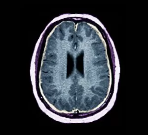
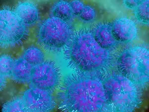
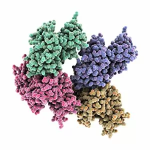
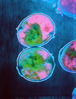
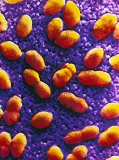
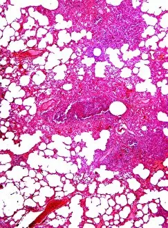
"Unveiling the Intricacies of Streptococcus pneumoniae: A Microscopic Journey into Bacterial Meningitis and Pneumonia" In this captivating journey through microscopic views, we explore the notorious bacterium known as Streptococcus pneumoniae. This pathogen is responsible for causing bacterial meningitis, a severe infection that affects the protective membranes surrounding the brain and spinal cord. Through an MRI scan, medical professionals can identify the presence of this bacterium in cases of suspected meningitis. The microscopic view reveals a diplococcus bacterium arrangement characteristic of S. Pneumoniae, resembling pairs of spherical cells. Not stopping at its involvement in meningitis alone, S. Pneumoniae also plays a significant role in bacterial pneumonia. Examining another microscopic view unveils clusters of these cocci bacteria invading lung tissues, causing inflammation and respiratory distress. Delving deeper into its structure, we observe distinct features such as corncob formations within dental plaque under examination with high magnification microscopy. These unique formations provide insights into how S. Pneumoniae adheres to surfaces and contributes to oral health complications. Furthermore, our exploration extends to specific enzymes produced by this bacterium – C3-degrading proteinase enzyme C016/1363 and C016/1362 – which aid in evading host immune responses by breaking down complement proteins involved in defense mechanisms. Streptococcus pneumoniae's ability to cause devastating infections like bacterial meningitis and contribute to widespread diseases like bacterial pneumonia highlights its significance within the realm of infectious diseases research. By unraveling its intricate nature through advanced imaging techniques, scientists strive towards developing effective prevention strategies and treatments against this formidable pathogen.