
Stimulus Collection
"Unveiling the Intricate Dance of Stimulus: From Phantom Pain to Artwork" In this captivating journey, we delve into the realm of stimulus


All Professionally Made to Order for Quick Shipping
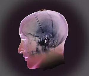
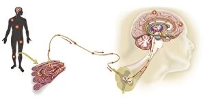
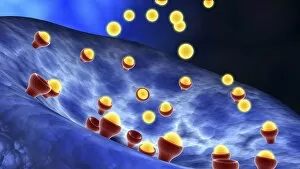
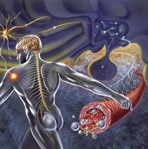
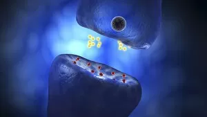
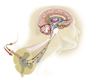
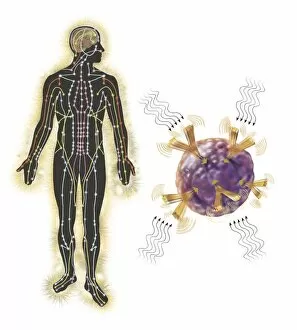
"Unveiling the Intricate Dance of Stimulus: From Phantom Pain to Artwork" In this captivating journey, we delve into the realm of stimulus, where a myriad of connections intertwine to shape our perception and experience. Like phantom pain after amputation, stimuli can evoke sensations that persist long after their physical presence has vanished. Plate II from Contributions of the physiology of vision No reveals an artistic depiction that mirrors how our senses influence our thoughts. Ivan Pavlov's caricature C013/7594 serves as a reminder that even renowned scientists were captivated by the enigma of stimulus. Follow the pathway of a pain message via sensory nerve in an injured muscle, and witness how it triggers a cascade within us. Our emotions are stirred as represented in artwork showcasing how our thoughts affect them. Conceptual images such as synaptic vesicles and nerve endings remind us of the intricate mechanisms at play. A nerve synapse demonstrates neurotransmitters' release, highlighting their role in transmitting signals between neurons. Observe synapse receptors through conceptual imagery, unveiling yet another layer in this complex web. Schematic representations reveal how the hypothalamus receives nerve impulses from throughout our body – orchestrating responses beyond conscious control. Finally, explore the human brain's sensory cortex layout – a mesmerizing map guiding us through various stimuli's processing centers. Stimulus is not merely an external force; it is an orchestration within ourselves – shaping perceptions, triggering emotions, and influencing every aspect of our being. Let these glimpses into its intricacies ignite curiosity about this profound phenomenon that defines so much of what it means to be human.