
Squamous Epithelium Collection
Squamous epithelium, a type of tissue found in various parts of the body, is an intriguing subject to explore


All Professionally Made to Order for Quick Shipping
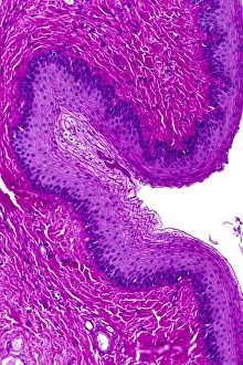
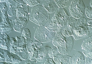
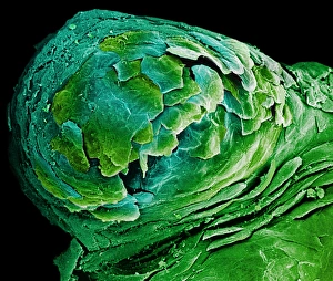
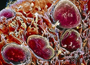
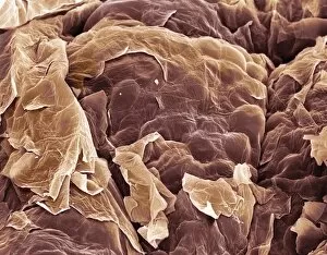
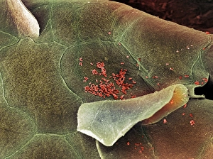
Squamous epithelium, a type of tissue found in various parts of the body, is an intriguing subject to explore. From cytologic smears of the vagina with intermediate squamous cells to conceptual images showcasing stratified squamous epithelium, this cellular structure offers a fascinating glimpse into our anatomy. One can visualize the oesophagus wall through light micrographs, revealing the intricate layers of stratified squamous epithelium that protect and line this vital organ. These microscopic images provide us with a closer look at the specialized cells that form this barrier against harmful substances. Confocal micrographs capture the beauty and complexity as well. The detailed image showcases its unique characteristics and highlights its role in different tissues such as endometrial or cervical areas. Squamous metaplasia is also observed in these regions, emphasizing how cellular transformations occur within our bodies. Even on our skin's surface, we find squamous epithelial cells forming a protective layer against external factors. Scanning electron microscopy allows us to appreciate their scale and arrangement, reminding us of their crucial function in safeguarding our bodies from potential harm. Exploring squamous epithelium through various imaging techniques provides valuable insights into its diverse forms across different anatomical locations. Whether it be examining vaginal cytology or visualizing metaplastic changes in cervical tissue, these captivating glimpses into cellular structures deepen our understanding of human biology and highlight the intricacies within ourselves.