
Spongy Bone Collection
"Spongy Bone: A Marvel of Structure and Strength" Delve into the intricate world with this captivating light micrograph, revealing its unique composition


All Professionally Made to Order for Quick Shipping
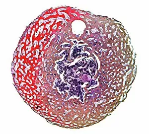
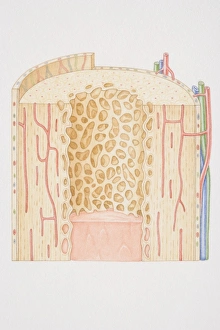
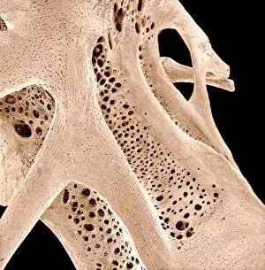
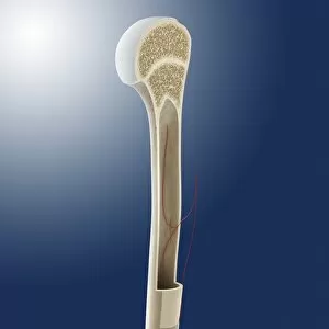
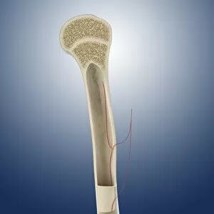
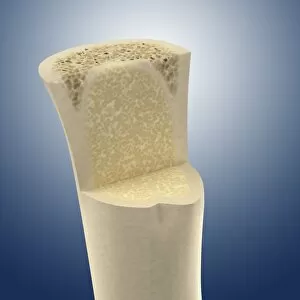
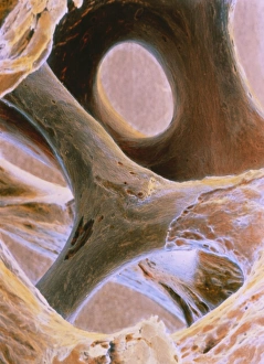
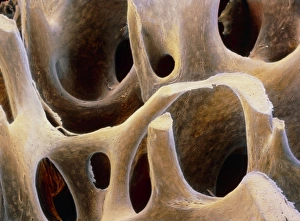
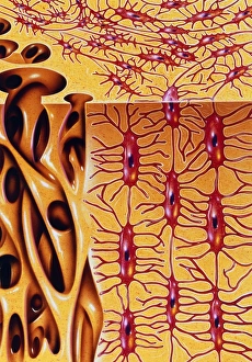
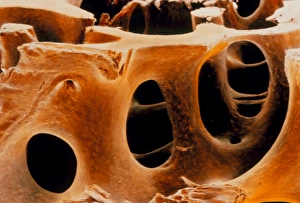
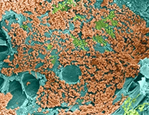
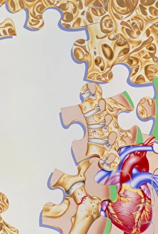
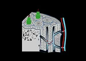
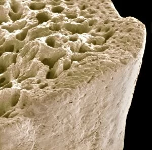
"Spongy Bone: A Marvel of Structure and Strength" Delve into the intricate world with this captivating light micrograph, revealing its unique composition. Explore the inner workings of a human long bone through a cross-section diagram, uncovering the fascinating intricacies of spongy bone. Witness the remarkable journey of childhood development in a biomedical illustration showcasing the cross section of a growing long bone. Behold the fully developed long bone in an adult as depicted in another biomedical illustration, highlighting the complexity and strength of spongy bone. Dive deeper into understanding the structure of human bones with an insightful biomedical illustration that unveils their hidden secrets. Immerse yourself in artistry as you admire artwork C016/7504, capturing the essence and beauty of bone structure like never before. Uncover nature's wonders with stunning artwork that showcases both fragility and resilience within our skeletal system. Get up close and personal with spongy bone tissue through scanning electron microscopy (SEM), revealing its intricate network and texture. Embark on an underwater adventure as SEM captures fish bones' delicate yet robust nature - behold SEM C015/8068. Discover more mesmerizing details from beneath ocean waves with SEM image C015/8070, showcasing fish bones' extraordinary architecture. Intricate, resilient, and awe-inspiring – spongy bone is truly a marvel worth exploring.