Fraud Blocker Skip to main content

Spinal Collection (page 7)

"Unlocking the Secrets of Spinal Harmony: Exploring Chakras and the Nervous System" Delve into the intricate world of the spinal column

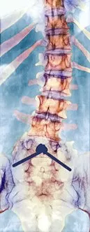
Background imageSpinal Collection: Pinned curved spine, X-ray

Pinned curved spine, X-ray
Pinned curved spine. Coloured X-ray of bones of the spine and pelvis of a 49 year old woman showing two screws (blue) pinning the lower lumbar vertebra (L5) to the sacrum of the pelvis

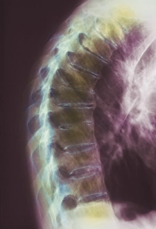
Background imageSpinal Collection: Osteoporotic spine, X-ray

Osteoporotic spine, X-ray
Osteoporotic spine, coloured X-ray. The spine is seen in side view, with the front of the body at right. It is seen to be curved, bowing out from top right to centre left then back right again

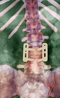
Background imageSpinal Collection: Pinned vertebrae, X-ray

Pinned vertebrae, X-ray
Pinned vertebrae. Coloured X-ray of pins (beige) joining two of the vertebrae (pink) of a 29 year old woman together to immobilise them. Vertebrae are the box-like bones that make up the spine

Background imageSpinal Collection: Slipped disc, CT scan

Slipped disc, CT scan
Slipped disc. Coloured computed tomography (CT) scan of an axial section through an inter- vertebral disc (orange) showing herniation

Background imageSpinal Collection: Osteoporosis of spine, X-ray

Osteoporosis of spine, X-ray
Osteoporosis. Coloured X-ray of a rear view of the lumbar (lower) spine of an elderly woman showing scoliosis (sideways curvature) of the spine caused by osteoporosis

Background imageSpinal Collection: Spinal osteoporosis

Spinal osteoporosis
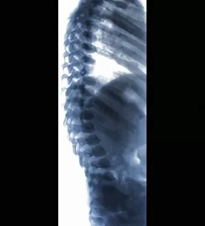
Curved spine in osteoporosis. Computer-enhanced X-ray of the curved and deformed thoracic spine of a woman suffering from osteoporosis

Background imageSpinal Collection: Osteoarthritis of neck, X-ray

Osteoarthritis of neck, X-ray
Osteoarthritis of the neck. Coloured X-ray of a side view of the neck of an elderly man, showing degenerative osteoarthritis of the cervical bones of the neck. Also known as cervical spondylosis

Background imageSpinal Collection: Ankylosing spondylitis X-ray

Ankylosing spondylitis X-ray
Ankylosing spondylitis. Coloured X-ray of the spine of a patient with ankylosing spondylitis, a form of arthritis. This is the lumbar region in the lower back

Background imageSpinal Collection: Scoliosis of the spine, artwork

Scoliosis of the spine, artwork

Background imageSpinal Collection: Vertebra, SEM

Vertebra, SEM
Vertebra. Coloured scanning electron micrograph (SEM) of a single vertebra, part of the vertebral column (spine). The spine is a column of 33 vertebrae (in humans)

Background imageSpinal Collection: Normal head and neck, MRI scan

Normal head and neck, MRI scan
Normal head and neck, coloured coronal magnetic resonance imaging (MRI) scan. The cervical (neck) spinal bones (vertebrae) are seen running from lower centre to upper centre

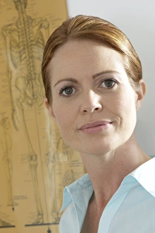
Background imageSpinal Collection: Chiropractor and patient

Chiropractor and patient
MODEL RELEASED. Chiropractor and patient. The chiropractor is using a model of the spine to the patients condition to her

Background imageSpinal Collection: Normal torso, CT scan

Normal torso, CT scan
Normal torso, coloured frontal computed tomography (CT) scan. This is a scout image showing the bones of the spine (vertebrae, centre)

Background imageSpinal Collection: Chiropractic examination

Chiropractic examination
MODEL RELEASED. Chiropractic examination of the thoracic spine, rib joints and lumbar spine

Background imageSpinal Collection: Normal neck, CT scan

Normal neck, CT scan
Normal neck, coloured coronal CT (computed tomography) scan. Spinal bones (cervical vertebrae) are light blue, and soft tissue is dark blue. These bones make up the top part of the spine

Background imageSpinal Collection: Chiropractor

Chiropractor
MODEL RELEASED. Chiropractor in her practice. Behind her is a chart including a diagram of the human skeleton. Chiropractic medicine is based on the theory that nearly all disorders can be traced to
Background imageSpinal Collection: Chiropractic treatment

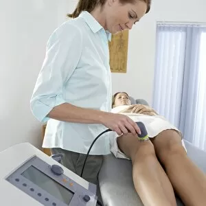
Chiropractic treatment
MODEL RELEASED. Chiropractic treatment. Chiropractor using electrotherapy to treat a complaint in her patients knee. The machine she is using combines ultrasound and electrotherapy

Background imageSpinal Collection: Lumbar spine and sacrum, computer artwork

Lumbar spine and sacrum, computer artwork
Lumbar spine and sacrum, side view, computer artwork. The bones of the spine (vertebrae) are blue and the intervertebral discs are red. The sacrum is at bottom

Background imageSpinal Collection: Lumbar spine, computer artwork

Lumbar spine, computer artwork
Lumbar spine, side view, computer artwork. The bones of the spine (vertebrae) are blue and the intervertebral discs are red

Background imageSpinal Collection: False-col X-ray of lumbar spine of woman

False-col X-ray of lumbar spine of woman
Diseased spine. Frontal coloured X-ray of the lumbar spine of a 60 year old suffering from scoliosis, osteophytosis and wasting of the spine

Background imageSpinal Collection: Pars vertebral injury, artwork

Pars vertebral injury, artwork
Artwork (side view) of the vertebrae of the spine showing a stress fracture (inset) of the pars interarticularis bone on lumbar 5 (L5) vertebra in the lower back

Background imageSpinal Collection: Fusion of spinal bones, X-ray

Fusion of spinal bones, X-ray
Coloured X-ray (front view) of the spine in a 45 year old patient with degenerative disc disease showing two implants (prostheses) between vertebral bones L4-L5 and L5-S1

Background imageSpinal Collection: Lumbar spine and sacrum, artwork

Lumbar spine and sacrum, artwork
Artwork of the five lumbar (lower back) vertebrae and sacrum (at bottom) of the spine. Bones are white, intervertebral discs are blue. The lower back is typically a site of back pain

Background imageSpinal Collection: Disability sexuality, conceptual artwork

Disability sexuality, conceptual artwork
Conceptual illustration showing sexual attraction between a man and a woman, both with physical disabilities

Background imageSpinal Collection: Spine in Morquio syndrome, X-ray

Spine in Morquio syndrome, X-ray
Coloured X-ray (side view) of the spine in a 7 year old patient with Morquio syndrome showing deformed vertebral bones (osteodysplasia). Morquio syndrome is an inherited lysosomal storage disorder

Background imageSpinal Collection: Bone density scanner, composite image

Bone density scanner, composite image
MODEL RELEASED. Bone density scanner. Composite image of a dual energy X-ray absorptiometry (DXA) scanner being used to measure a patients bone mineral density (BMD)

Background imageSpinal Collection: Chest X-ray imaging, composite image

Chest X-ray imaging, composite image. At left a ceiling-mounted X-ray machine is being used to X-ray a patients chest. The artwork of the patient (at right)

Background imageSpinal Collection: Correct lifting technique, artwork

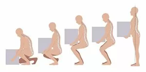
Correct lifting technique, artwork
Correct lifting technique. Artwork showing the correct posture to use when lifting heavy or bulky items. All the bending should be done at the knees, leaving the back straight

Background imageSpinal Collection: Main in pain, artwork

Main in pain, artwork
Main in pain, computer artwork

Background imageSpinal Collection: Pregnancy and gravity, artwork

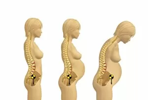
Pregnancy and gravity, artwork
Pregnancy and gravity. Computer artwork showing how increasing body mass on the front of a womans body during pregnancy - caused by enlarged breasts

Background imageSpinal Collection: Calcified aortic artery, 3D CT scan

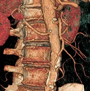
Calcified aortic artery, 3D CT scan
Coloured three-dimensional (3D) Computed Tomography (CT) scan in front view of the abdomen in a patient showing the kidneys (red)

Background imageSpinal Collection: Spine and aorta, 3D CT scan

Spine and aorta, 3D CT scan
Coloured three-dimensional (3D) Computed Tomography (CT) scan in front view of the abdomen in a patient showing the vertebral bones of the spine (running top to bottom)

Background imageSpinal Collection: Heart and aortic artery, 3D CT scan

Heart and aortic artery, 3D CT scan
Coloured three-dimensional (3D) Computed Tomography (CT) scan in side view of the vertebral bones (spine, at left) in a patient showing the heart (at top right)

Background imageSpinal Collection: Slipped disc, MRI scan

Slipped disc, MRI scan
Slipped disc, coloured magnetic resonance imaging (MRI) scan. Profile view of the spine of a 59-year-old patient with severe rheumatoid arthritis and a herniated (slipped) disc (red)

Background imageSpinal Collection: Kidneys and bladder, 3D CT scan

Kidneys and bladder, 3D CT scan
Coloured three-dimensional (3D) Computed Tomography (CT) scan in front view of the abdomen in a patient showing the kidneys (blue) on either side of the vertebral bones of the spine (at upper centre)

Background imageSpinal Collection: Spine in Hurler Syndrome, X-ray

Spine in Hurler Syndrome, X-ray
Spine in Hurler Syndrome. Coloured X-ray of the spine of a 26-month-old child with Hurler syndrome (also called mucopolysaccharidosis type I, MPS I)


All Professionally Made to Order for Quick Shipping

"Unlocking the Secrets of Spinal Harmony: Exploring Chakras and the Nervous System" Delve into the intricate world of the spinal column, where ancient wisdom meets modern science. Discover how this remarkable structure, revered by pioneers like Ramon y Cajal, holds the key to our physical and spiritual well-being. Embark on a visual journey through time as you explore a diagram showcasing the human brain and spinal cord. Witness how these interconnected powerhouses orchestrate every aspect of our existence, from bodily functions to emotional experiences. Step back in time with Calots spinal surgery, an innovation from the 19th century that revolutionized medical practices. Marvel at illustrations depicting this groundbreaking procedure that aimed to alleviate pain and restore mobility. Peer into X-ray images capturing both normal necks and spines, revealing their intricate beauty hidden beneath our skin. Observe an extended neck vertebrae X-ray, shedding light on potential issues caused by misalignment or injury. Art comes alive as body pain is transformed into captivating artwork – a poignant reminder of the challenges faced by those afflicted with discomfort along their spine. Let your imagination soar as upper body skeletons take form through computer-generated artistry. Witness how lower back pain manifests itself in conceptual artwork – a vivid representation of its impact on daily life. Explore similar expressions for upper back pain; each stroke conveying tales of struggle yet resilience against this common ailment. Intriguingly juxtaposing these artistic interpretations are X-rays showcasing normal necks and spines - silent testaments to health and balance within our bodies' core structures. Uncover new perspectives on spinal health as you immerse yourself in this captivating collection.