
Spinal Nerves Collection
"Exploring the Intricacies of Spinal Nerves: Unveiling the Wonders Within" Delving into the depths of human anatomy


All Professionally Made to Order for Quick Shipping
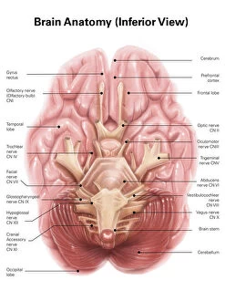
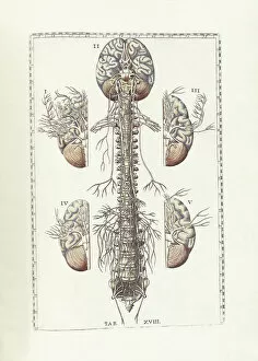
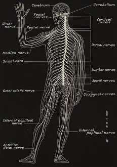
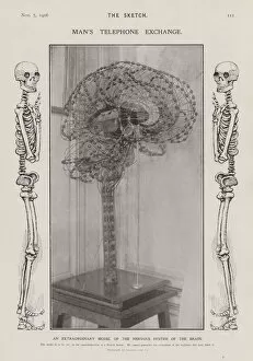
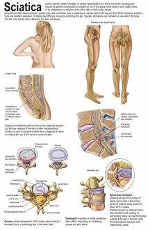
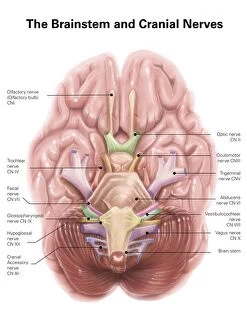
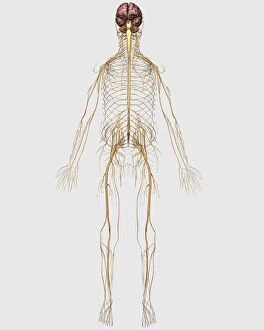
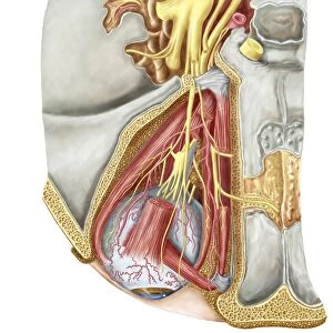
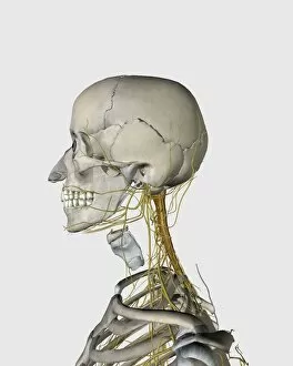
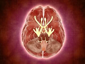
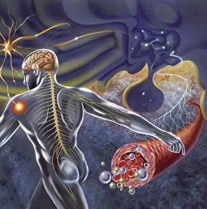
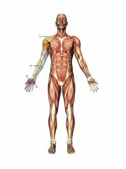
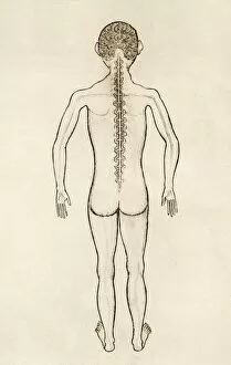
"Exploring the Intricacies of Spinal Nerves: Unveiling the Wonders Within" Delving into the depths of human anatomy, an inferior view of the brain reveals a complex network known as spinal nerves. As depicted in Bartholomeo Eustachi's masterpiece, "The Science of Human Anatomy, " these delicate structures play a vital role in our body's functioning. Dating back to 1844, an exquisite artwork showcases cervical spinal nerves, capturing their intricate details and highlighting their significance. Santiago Ramon y Cajal, a renowned Spanish histologist and Nobel laureate in medicine, further immortalized these nerves with his vibrant colored drawing titled "Shema histologique des nerfs de la colonne vertebrale. " Lithographs such as "Spinal Cord and Spinal Nerves" provide us with visual insights into this crucial system. Coupled with a black-and-white photograph showcasing a model of the nervous system within the human brain, we gain a deeper understanding of its complexity. Medical illustrations serve as valuable tools for education and diagnosis. From detailing thoracic outlet syndrome to portraying sciatica caused by herniated discs, they shed light on conditions that can affect our spinal nerves. The peripheral nervous system is intricately connected to our brain. A medical illustration beautifully captures this relationship while emphasizing its importance in maintaining bodily functions. Intriguingly, an orbital cut unravels yet another aspect – unveiling the abducent nerve alongside the ciliary ganglion and oculomotor nerve. This glimpse into ocular anatomy highlights how spinal nerves extend beyond mere bodily movements but also contribute to sensory perception. As we explore each facet captured through artistry or scientific documentation like anatomical drawings or medical illustrations depicting various aspects related to spinal nerves - from their origin within vertebral bones to their impact on peripheral systems - we come closer to comprehending their immense significance.